 Movie
Movie Controller

[English] 日本語
 Yorodumi
- PDB-2om7: Structural Basis for Interaction of the Ribosome with the Switch ... -
+ Open data

- Basic information
| Entry | Database: PDB / ID: 2om7 | ||||||
|---|---|---|---|---|---|---|---|
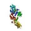
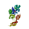
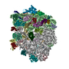
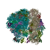
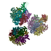
| Title | Structural Basis for Interaction of the Ribosome with the Switch Regions of GTP-bound Elongation Factors | ||||||
 Components Components |
| ||||||
 Keywords Keywords | RIBOSOME / RNA-Protein Complex | ||||||
| Function / homology |  Function and homology information Function and homology informationribosome disassembly / translational elongation / translation elongation factor activity / GDP binding / regulation of translation / large ribosomal subunit / ribosome binding / small ribosomal subunit / cytosolic small ribosomal subunit / tRNA binding ...ribosome disassembly / translational elongation / translation elongation factor activity / GDP binding / regulation of translation / large ribosomal subunit / ribosome binding / small ribosomal subunit / cytosolic small ribosomal subunit / tRNA binding / rRNA binding / structural constituent of ribosome / translation / response to antibiotic / GTPase activity / GTP binding / magnesium ion binding / cytoplasm Similarity search - Function | ||||||
| Biological species |   Thermus thermophilus (bacteria) Thermus thermophilus (bacteria) | ||||||
| Method | ELECTRON MICROSCOPY / single particle reconstruction / cryo EM / Resolution: 7.3 Å | ||||||
 Authors Authors | Connell, S.R. / Wilson, D.N. / Rost, M. / Schueler, M. / Giesebrecht, J. / Dabrowski, M. / Mielke, T. / Fucini, P. / Spahn, C.M.T. | ||||||
 Citation Citation | Journal: Mol Cell / Year: 2007 Title: Structural basis for interaction of the ribosome with the switch regions of GTP-bound elongation factors. Authors: Sean R Connell / Chie Takemoto / Daniel N Wilson / Hongfei Wang / Kazutaka Murayama / Takaho Terada / Mikako Shirouzu / Maximilian Rost / Martin Schüler / Jan Giesebrecht / Marylena ...Authors: Sean R Connell / Chie Takemoto / Daniel N Wilson / Hongfei Wang / Kazutaka Murayama / Takaho Terada / Mikako Shirouzu / Maximilian Rost / Martin Schüler / Jan Giesebrecht / Marylena Dabrowski / Thorsten Mielke / Paola Fucini / Shigeyuki Yokoyama / Christian M T Spahn /  Abstract: Elongation factor G (EF-G) catalyzes tRNA translocation on the ribosome. Here a cryo-EM reconstruction of the 70S*EF-G ribosomal complex at 7.3 A resolution and the crystal structure of EF-G-2*GTP, ...Elongation factor G (EF-G) catalyzes tRNA translocation on the ribosome. Here a cryo-EM reconstruction of the 70S*EF-G ribosomal complex at 7.3 A resolution and the crystal structure of EF-G-2*GTP, an EF-G homolog, at 2.2 A resolution are presented. EF-G-2*GTP is structurally distinct from previous EF-G structures, and in the context of the cryo-EM structure, the conformational changes are associated with ribosome binding and activation of the GTP binding pocket. The P loop and switch II approach A2660-A2662 in helix 95 of the 23S rRNA, indicating an important role for these conserved bases. Furthermore, the ordering of the functionally important switch I and II regions, which interact with the bound GTP, is dependent on interactions with the ribosome in the ratcheted conformation. Therefore, a network of interaction with the ribosome establishes the active GTP conformation of EF-G and thus facilitates GTP hydrolysis and tRNA translocation. | ||||||
| History |
|
- Structure visualization
| Movie |
Movie viewer |
|---|---|
| Structure viewer | Molecule: MolmilJmol/JSmol |
- Downloads & links
-Download
| PDBx/mmCIF format | 2om7.cif.gz | 459 KB | Display | PDBx/mmCIF format |
|---|---|---|---|---|
| PDB format | pdb2om7.ent.gz | 331.4 KB | Display | PDB format |
| PDBx/mmJSON format | 2om7.json.gz | Tree view | PDBx/mmJSON format | |
| Others |  Other downloads Other downloads |
-Validation report
| Arichive directory | https://data.pdbj.org/pub/pdb/validation_reports/om/2om7ftp://data.pdbj.org/pub/pdb/validation_reports/om/2om7 | HTTPS FTP |
|---|
-Related structure data
| Related structure data | 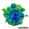 1315MC M: map data used to model this data C: citing same article ( |
|---|---|
| Similar structure data |


-Links
 PDBj
PDBj


- Assembly
| Deposited unit | 
|
|---|---|
| 1 |
|
| Details | A model for the entire complex can be generated by aligning the 50s subunit, 30s Head and 30s body from PDB IDs 2j00 and 2j01 to the corresponding elements in this model |
-Components
-Fragment of 16S rRNA ... , 3 types, 3 molecules ABC
| #1: RNA chain | Mass: 3827.336 Da / Num. of mol.: 1 / Source method: isolated from a natural source / Details: 16S rRNA / Source: (natural) Thermus thermophilus (bacteria) |
|---|---|
| #2: RNA chain | Mass: 9048.452 Da / Num. of mol.: 1 / Source method: isolated from a natural source / Details: 16S rRNA / Source: (natural) Thermus thermophilus (bacteria) |
| #3: RNA chain | Mass: 31208.635 Da / Num. of mol.: 1 / Source method: isolated from a natural source / Details: 16S rRNA / Source: (natural) Thermus thermophilus (bacteria) / References: GenBank: 118505352 |
-RNA chain , 2 types, 2 molecules DM
| #4: RNA chain | Mass: 98292.133 Da / Num. of mol.: 1 / Source method: isolated from a natural source / Details: 16S rRNA / Source: (natural) Thermus thermophilus (bacteria) / References: GenBank: 48256 |
|---|---|
| #10: RNA chain | Mass: 23728.123 Da / Num. of mol.: 1 / Source method: isolated from a natural source / Details: tRNA / Source: (natural) Thermus thermophilus (bacteria) |
-Fragment of23S rRNA ... , 5 types, 5 molecules FGHIJ
| #5: RNA chain | Mass: 9378.643 Da / Num. of mol.: 1 / Source method: isolated from a natural source / Details: 23S rRNA / Source: (natural) Thermus thermophilus (bacteria) |
|---|---|
| #6: RNA chain | Mass: 17631.637 Da / Num. of mol.: 1 / Source method: isolated from a natural source / Details: 23S rRNA / Source: (natural) Thermus thermophilus (bacteria) / References: GenBank: 37223182 |
| #7: RNA chain | Mass: 13540.067 Da / Num. of mol.: 1 / Source method: isolated from a natural source / Details: 23S rRNA / Source: (natural) Thermus thermophilus (bacteria) |
| #8: RNA chain | Mass: 18710.129 Da / Num. of mol.: 1 / Source method: isolated from a natural source / Details: 23S rRNA / Source: (natural) Thermus thermophilus (bacteria) / References: GenBank: 37223182 |
| #9: RNA chain | Mass: 33148.680 Da / Num. of mol.: 1 / Source method: isolated from a natural source / Details: 23S rRNA / Source: (natural) Thermus thermophilus (bacteria) / References: GenBank: 37223182 |
-30S ribosomal protein ... , 2 types, 2 molecules EN
| #11: Protein | Mass: 14920.754 Da / Num. of mol.: 1 / Source method: isolated from a natural source / Details: Small ribosomal subunit protein S12 / Source: (natural) Thermus thermophilus (bacteria) / References: UniProt: P17293, UniProt: Q5SHN3*PLUS |
|---|---|
| #14: Protein | Mass: 29317.703 Da / Num. of mol.: 1 / Source method: isolated from a natural source / Details: Small ribosomal subunit protein S2 / Source: (natural) Thermus thermophilus (bacteria) / References: UniProt: P80371 |
-Protein , 2 types, 2 molecules KL
| #12: Protein | Mass: 24872.721 Da / Num. of mol.: 1 / Source method: isolated from a natural source / Details: Large ribosomal subunit protein L1 / Source: (natural) Thermus thermophilus (bacteria) / References: UniProt: Q5SLP7 |
|---|---|
| #13: Protein | Mass: 76977.102 Da / Num. of mol.: 1 Source method: isolated from a genetically manipulated source Source: (gene. exp.) Thermus thermophilus (bacteria) / Gene: fusA, fus / Species (production host): Escherichia coli / Production host: |
-Experimental details
-Experiment
| Experiment | Method: ELECTRON MICROSCOPY |
|---|---|
| EM experiment | Aggregation state: PARTICLE / 3D reconstruction method: single particle reconstruction |
- Sample preparation
| Component |
| ||||||||||||||||||||||||
|---|---|---|---|---|---|---|---|---|---|---|---|---|---|---|---|---|---|---|---|---|---|---|---|---|---|
| Buffer solution | Name: 0.3 mM GMPPNP, 10 mM Hepes-KOH (pH 7.8), 10 mM Mg acetate, 60 mM NH4Cl, and 6 mM B-mercaptoethanol pH: 7.8 Details: 0.3 mM GMPPNP, 10 mM Hepes-KOH (pH 7.8), 10 mM Mg acetate, 60 mM NH4Cl, and 6 mM B-mercaptoethanol | ||||||||||||||||||||||||
| Specimen | Embedding applied: NO / Shadowing applied: NO / Staining applied: NO / Vitrification applied: YES | ||||||||||||||||||||||||
| Vitrification | Instrument: FEI VITROBOT MARK I / Cryogen name: ETHANE / Humidity: 95 % / Chamber temperature: 277 K |
- Electron microscopy imaging
| Experimental equipment |  Model: Tecnai F30 / Image courtesy: FEI Company |
|---|---|
| Microscopy | Model: FEI TECNAI F30 |
| Electron gun | Electron source:  FIELD EMISSION GUN / Accelerating voltage: 300 kV / Illumination mode: FLOOD BEAM FIELD EMISSION GUN / Accelerating voltage: 300 kV / Illumination mode: FLOOD BEAM |
| Electron lens | Mode: BRIGHT FIELD / Nominal magnification: 39000 X / Nominal defocus max: 4000 nm / Nominal defocus min: 800 nm |
| Specimen holder | Specimen holder type: Eucentric |
| Image recording | Electron dose: 19 e/Å2 / Film or detector model: KODAK SO-163 FILM |
- Processing
| Software | Name: SPIDER / Classification: refinement | ||||||||||||||||||||||||||||||||||||||||||
|---|---|---|---|---|---|---|---|---|---|---|---|---|---|---|---|---|---|---|---|---|---|---|---|---|---|---|---|---|---|---|---|---|---|---|---|---|---|---|---|---|---|---|---|
| EM software |
| ||||||||||||||||||||||||||||||||||||||||||
| CTF correction | Details: defocus groups | ||||||||||||||||||||||||||||||||||||||||||
| Symmetry | Point symmetry: C1 (asymmetric) | ||||||||||||||||||||||||||||||||||||||||||
| 3D reconstruction | Resolution: 7.3 Å / Num. of particles: 77038 Details: The geometry of linkages between some residues in chains J, M and L are distorted. They were not resolved based on the data used for solving this structure. Symmetry type: POINT | ||||||||||||||||||||||||||||||||||||||||||
| Atomic model building | Protocol: RIGID BODY FIT / Details: METHOD--Rigid Body | ||||||||||||||||||||||||||||||||||||||||||
| Atomic model building |
| ||||||||||||||||||||||||||||||||||||||||||
| Refinement step | Cycle: LAST
|

