 Movie
Movie Controller

[English] 日本語
 Yorodumi
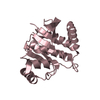
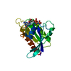
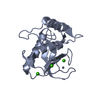
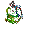
- PDB-2oh1: Crystal structure of acetyltransferase GNAT family (YP_013287.1) ... -
+ Open data

- Basic information
| Entry | Database: PDB / ID: 2oh1 | ||||||
|---|---|---|---|---|---|---|---|
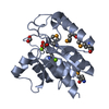
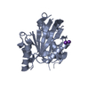
| Title | Crystal structure of acetyltransferase GNAT family (YP_013287.1) from Listeria monocytogenes 4b F2365 at 1.46 A resolution | ||||||
 Components Components | Acetyltransferase, GNAT family | ||||||
 Keywords Keywords | TRANSFERASE / YP_013287.1 / acetyltransferase GNAT family / Structural Genomics / Joint Center for Structural Genomics / JCSG / Protein Structure Initiative / PSI-2 | ||||||
| Function / homology | Gcn5-related N-acetyltransferase (GNAT) / Aminopeptidase / 3-Layer(aba) Sandwich / Alpha Beta / Unknown ligand / :  Function and homology information Function and homology information | ||||||
| Biological species |  Listeria monocytogenes str. 4b (bacteria) Listeria monocytogenes str. 4b (bacteria) | ||||||
| Method |  X-RAY DIFFRACTION / SYNCHROTRON / MAD / Resolution: 1.46 Å X-RAY DIFFRACTION / SYNCHROTRON / MAD / Resolution: 1.46 Å | ||||||
 Authors Authors | Joint Center for Structural Genomics (JCSG) | ||||||
 Citation Citation | Journal: To be published Title: Crystal structure of acetyltransferase GNAT family (YP_013287.1) from Listeria monocytogenes 4b F2365 at 1.46 A resolution Authors: Joint Center for Structural Genomics (JCSG) | ||||||
| History |
| ||||||
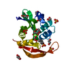
| Remark 300 | BIOMOLECULE: 1,2,3,4 THIS ENTRY CONTAINS THE CRYSTALLOGRAPHIC ASYMMETRIC UNIT WHICH CONSISTS OF 4 ... BIOMOLECULE: 1,2,3,4 THIS ENTRY CONTAINS THE CRYSTALLOGRAPHIC ASYMMETRIC UNIT WHICH CONSISTS OF 4 CHAIN(S). SEE REMARK 350 FOR INFORMATION ON GENERATING THE BIOLOGICAL MOLECULE(S). AUTHORS STATE THAT THE STATIC LIGHT SCATTERING WITH ANALYTICAL SIZE EXCLUSION CHROMATOGRAPHY MEASUREMENTS INDICATE THAT THE MONOMER IS A BIOLOGICALLY SIGNIFICANT OLIGOMERIZATION STATE IN SOLUTION. | ||||||
| Remark 999 | SEQUENCE THE CONSTRUCT WAS EXPRESSED WITH A PURIFICATION TAG MGSDKIHHHHHHENLYFQG. THE TAG WAS ... SEQUENCE THE CONSTRUCT WAS EXPRESSED WITH A PURIFICATION TAG MGSDKIHHHHHHENLYFQG. THE TAG WAS REMOVED WITH TEV PROTEASE LEAVING ONLY A GLYCINE (SEQUENCE NUMBER 0) FOLLOWED BY THE TARGET SEQUENCE. |
- Structure visualization
| Structure viewer | Molecule: MolmilJmol/JSmol |
|---|
- Downloads & links
-Download
| PDBx/mmCIF format | 2oh1.cif.gz | 179.9 KB | Display | PDBx/mmCIF format |
|---|---|---|---|---|
| PDB format | pdb2oh1.ent.gz | 144.8 KB | Display | PDB format |
| PDBx/mmJSON format | 2oh1.json.gz | Tree view | PDBx/mmJSON format | |
| Others |  Other downloads Other downloads |
-Validation report
| Arichive directory | https://data.pdbj.org/pub/pdb/validation_reports/oh/2oh1ftp://data.pdbj.org/pub/pdb/validation_reports/oh/2oh1 | HTTPS FTP |
|---|
-Related structure data
| Similar structure data | |
|---|---|
| Other databases |


-Links
 PDBj
PDBj

- Assembly
| Deposited unit | 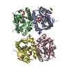
| ||||||||
|---|---|---|---|---|---|---|---|---|---|
| 1 | 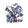
| ||||||||
| 2 | 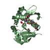
| ||||||||
| 3 | 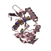
| ||||||||
| 4 | 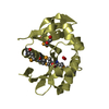
| ||||||||
| Unit cell |
| ||||||||
| Details | SIZE EXCLUSION CHROMATOGRAPHY SUPPORTS THE ASSIGNMENT OF A MONOMER AS A BIOLOGICALLY SIGNIFICANT OLIGOMERIZATION STATE. |
-Components
| #1: Protein | Mass: 20884.744 Da / Num. of mol.: 4 Source method: isolated from a genetically manipulated source Source: (gene. exp.) Listeria monocytogenes str. 4b (bacteria)Species: Listeria monocytogenes / Strain: F2365 / Gene: YP_013287.1, LMOf2365_0683 / Plasmid: SpeedET / Production host: #2: Chemical | Num. of mol.: 3 / Source method: obtained synthetically #3: Chemical | ChemComp-EDO /   Mass: 62.068 Da / Num. of mol.: 7 / Source method: obtained synthetically / Formula: C2H6O2 Mass: 62.068 Da / Num. of mol.: 7 / Source method: obtained synthetically / Formula: C2H6O2#4: Water | ChemComp-HOH / |  Mass: 18.015 Da / Num. of mol.: 838 / Source method: isolated from a natural source / Formula: H2O Mass: 18.015 Da / Num. of mol.: 838 / Source method: isolated from a natural source / Formula: H2OHas protein modification | Y | |
|---|
-Experimental details
-Experiment
| Experiment | Method: X-RAY DIFFRACTION / Number of used crystals: 1 |
|---|
- Sample preparation
| Crystal | Density Matthews: 2.41 Å3/Da / Density % sol: 48.94 % |
|---|---|
| Crystal grow | Temperature: 277 K / pH: 7 Details: NANODROP, 1.0M Na Citrate, 0.2M NaCl, 0.1M TRIS pH 7.0, VAPOR DIFFUSION, SITTING DROP, temperature 277K, pH 7.00 |
-Data collection
| Diffraction | Mean temperature: 100 K | ||||||||||||
|---|---|---|---|---|---|---|---|---|---|---|---|---|---|
| Diffraction source | Source: SYNCHROTRON / Site: SSRL  / Beamline: BL11-1 / Wavelength: 0.91837, 0.97932, 0.97901 / Beamline: BL11-1 / Wavelength: 0.91837, 0.97932, 0.97901 | ||||||||||||
| Detector | Type: MARMOSAIC 325 mm CCD / Detector: CCD / Date: Dec 3, 2006 / Details: FLAT MIRROR (VERTICAL FOCUSING) | ||||||||||||
| Radiation | Monochromator: SINGLE CRYSTAL SI(111) BENT (HORIZONTAL FOCUSING) Protocol: MAD / Monochromatic (M) / Laue (L): M / Scattering type: x-ray | ||||||||||||
| Radiation wavelength |
| ||||||||||||
| Reflection | Resolution: 1.46→46.225 Å / Num. obs: 125870 / % possible obs: 89.6 % / Biso Wilson estimate: 25.14 Å2 / Rmerge(I) obs: 0.055 / Net I/σ(I): 12.54 | ||||||||||||
| Reflection shell | Resolution: 1.46→1.51 Å / Rmerge(I) obs: 0.671 / Mean I/σ(I) obs: 1.79 / % possible all: 52.5 |
-Phasing
| Phasing | Method: MAD |
|---|
- Processing
| Software |
| |||||||||||||||||||||||||||||||||||||||||||||||||||||||||||||||||||||||||||||||||||||||||||||||||||||||||||||||||||||||||||||
|---|---|---|---|---|---|---|---|---|---|---|---|---|---|---|---|---|---|---|---|---|---|---|---|---|---|---|---|---|---|---|---|---|---|---|---|---|---|---|---|---|---|---|---|---|---|---|---|---|---|---|---|---|---|---|---|---|---|---|---|---|---|---|---|---|---|---|---|---|---|---|---|---|---|---|---|---|---|---|---|---|---|---|---|---|---|---|---|---|---|---|---|---|---|---|---|---|---|---|---|---|---|---|---|---|---|---|---|---|---|---|---|---|---|---|---|---|---|---|---|---|---|---|---|---|---|---|
| Refinement | Method to determine structure: MAD / Resolution: 1.46→46.225 Å / Cor.coef. Fo:Fc: 0.971 / Cor.coef. Fo:Fc free: 0.96 / SU B: 2.672 / SU ML: 0.05 / TLS residual ADP flag: LIKELY RESIDUAL / Cross valid method: THROUGHOUT / σ(F): 0 / ESU R: 0.071 / ESU R Free: 0.071 Stereochemistry target values: MAXIMUM LIKELIHOOD WITH PHASES Details: 1. HYDROGENS HAVE BEEN ADDED IN THE RIDING POSITIONS. 2. A MET-INHIBITION PROTOCOL WAS USED FOR SELENOMETHIONINE INCORPORATION DURING PROTEIN EXPRESSION. THE OCCUPANCY OF THE SE ATOMS IN THE ...Details: 1. HYDROGENS HAVE BEEN ADDED IN THE RIDING POSITIONS. 2. A MET-INHIBITION PROTOCOL WAS USED FOR SELENOMETHIONINE INCORPORATION DURING PROTEIN EXPRESSION. THE OCCUPANCY OF THE SE ATOMS IN THE MSE RESIDUES WAS REDUCED TO 0.75 TO ACCOUNT FOR THE REDUCED SCATTERING POWER DUE TO PARTIAL S-MET INCORPORATION. 3. ATOM RECORD CONTAINS RESIDUAL B FACTORS ONLY. 4. UNIDENTIFIED ELECTRON DENSITY FOUND NEAR THE ACTIVE SITE WAS MODELED AS AN UNKNOWN LIGAND (UNL). 5. THE ELECTRON DENSITIES SHOW THAT RESIDUES SER42 AND THR43 IN SUBUNIT A ARE DISORDERED, AND THESE RESIDUES WERE MODELED BASED ON THE MODEL FOR THE NCS-RELATED B SUBUNIT.
| |||||||||||||||||||||||||||||||||||||||||||||||||||||||||||||||||||||||||||||||||||||||||||||||||||||||||||||||||||||||||||||
| Solvent computation | Ion probe radii: 0.8 Å / Shrinkage radii: 0.8 Å / VDW probe radii: 1.2 Å / Solvent model: MASK | |||||||||||||||||||||||||||||||||||||||||||||||||||||||||||||||||||||||||||||||||||||||||||||||||||||||||||||||||||||||||||||
| Displacement parameters | Biso mean: 17.53 Å2
| |||||||||||||||||||||||||||||||||||||||||||||||||||||||||||||||||||||||||||||||||||||||||||||||||||||||||||||||||||||||||||||
| Refinement step | Cycle: LAST / Resolution: 1.46→46.225 Å
| |||||||||||||||||||||||||||||||||||||||||||||||||||||||||||||||||||||||||||||||||||||||||||||||||||||||||||||||||||||||||||||
| Refine LS restraints |
| |||||||||||||||||||||||||||||||||||||||||||||||||||||||||||||||||||||||||||||||||||||||||||||||||||||||||||||||||||||||||||||
| LS refinement shell | Resolution: 1.46→1.498 Å / Total num. of bins used: 20
| |||||||||||||||||||||||||||||||||||||||||||||||||||||||||||||||||||||||||||||||||||||||||||||||||||||||||||||||||||||||||||||
| Refinement TLS params. | Method: refined / Refine-ID: X-RAY DIFFRACTION
| |||||||||||||||||||||||||||||||||||||||||||||||||||||||||||||||||||||||||||||||||||||||||||||||||||||||||||||||||||||||||||||
| Refinement TLS group | Refine-ID: X-RAY DIFFRACTION / Selection: ALL
|
