 Movie
Movie Controller

[English] 日本語
 Yorodumi
- PDB-2obk: X-Ray structure of the putative Se binding protein from Pseudomon... -
+ Open data

- Basic information
| Entry | Database: PDB / ID: 2obk | ||||||
|---|---|---|---|---|---|---|---|
| Title | X-Ray structure of the putative Se binding protein from Pseudomonas fluorescens. Northeast Structural Genomics Consortium target PlR6. | ||||||
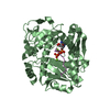
 Components Components | SelT/selW/selH selenoprotein domain | ||||||
 Keywords Keywords | STRUCTURAL GENOMICS / UNKNOWN FUNCTION / X-Ray NESG PlR6 Q4KGC5 / PSI-2 / Protein Structure Initiative / Northeast Structural Genomics Consortium | ||||||
| Function / homology | Selenoprotein, Rdx-type / Rdx family / Glutaredoxin / Glutaredoxin / Thioredoxin-like superfamily / 3-Layer(aba) Sandwich / Alpha Beta / SelT/selW/selH selenoprotein domain protein Function and homology information Function and homology information | ||||||
| Biological species |  Pseudomonas fluorescens (bacteria) Pseudomonas fluorescens (bacteria) | ||||||
| Method |  X-RAY DIFFRACTION / SYNCHROTRON / MOLECULAR REPLACEMENT / Resolution: 2.7 Å X-RAY DIFFRACTION / SYNCHROTRON / MOLECULAR REPLACEMENT / Resolution: 2.7 Å | ||||||
 Authors Authors | Kuzin, A.P. / Su, M. / Seetharaman, J. / Chen, C.X. / Fang, Y. / Cunningham, K. / Ma, L.C. / Xiao, R. / Liu, J. / Baran, M.C. ...Kuzin, A.P. / Su, M. / Seetharaman, J. / Chen, C.X. / Fang, Y. / Cunningham, K. / Ma, L.C. / Xiao, R. / Liu, J. / Baran, M.C. / Acton, T.B. / Rost, B. / Montelione, G.T. / Tong, L. / Hunt, J.F. / Northeast Structural Genomics Consortium (NESG) | ||||||
 Citation Citation | Journal: To be Published Title: X-Ray structure of the putative Se binding protein from Pseudomonas fluorescens Authors: Kuzin, A.P. / Su, M. / Seetharaman, J. / Chen, C. / Fang, Y. / Cunningham, K. / Ma, L.C. / Xiao, R. / Liu, J. / Baran, M.C. / Acton, T.B. / Rost, B. / Montelione, G.T. / Tong, L. / Hunt, J.F. | ||||||
| History |
|
- Structure visualization
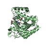
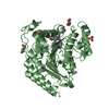
| Structure viewer | Molecule: MolmilJmol/JSmol |
|---|
- Downloads & links
-Download
| PDBx/mmCIF format | 2obk.cif.gz | 143.2 KB | Display | PDBx/mmCIF format |
|---|---|---|---|---|
| PDB format | pdb2obk.ent.gz | 113.9 KB | Display | PDB format |
| PDBx/mmJSON format | 2obk.json.gz | Tree view | PDBx/mmJSON format | |
| Others |  Other downloads Other downloads |
-Validation report
| Summary document | 2obk_validation.pdf.gz | 493.9 KB | Display | wwPDB validaton report |
|---|---|---|---|---|
| Full document | 2obk_full_validation.pdf.gz | 512 KB | Display | |
| Data in XML | 2obk_validation.xml.gz | 27.3 KB | Display | |
| Data in CIF | 2obk_validation.cif.gz | 38.2 KB | Display | |
| Arichive directory | https://data.pdbj.org/pub/pdb/validation_reports/ob/2obkftp://data.pdbj.org/pub/pdb/validation_reports/ob/2obk | HTTPS FTP |
-Related structure data
| Related structure data |  2fa8S S: Starting model for refinement |
|---|---|
| Similar structure data | |
| Other databases |


-Links
 PDBj
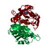
PDBj- Assembly
| Deposited unit | 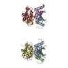
| ||||||||
|---|---|---|---|---|---|---|---|---|---|
| 1 | 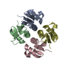
| ||||||||
| 2 | 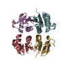
| ||||||||
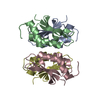
| Unit cell |
|
-Components
| #1: Protein | Mass: 11951.298 Da / Num. of mol.: 8 Source method: isolated from a genetically manipulated source Source: (gene. exp.) Pseudomonas fluorescens (bacteria) / Strain: Pf-5 / Gene: PFL_1582 / Plasmid: pET21 / Production host: #2: Water | ChemComp-HOH / |  Mass: 18.015 Da / Num. of mol.: 132 / Source method: isolated from a natural source / Formula: H2O Mass: 18.015 Da / Num. of mol.: 132 / Source method: isolated from a natural source / Formula: H2OHas protein modification | Y | |
|---|
-Experimental details
-Experiment
| Experiment | Method: X-RAY DIFFRACTION / Number of used crystals: 1 |
|---|
- Sample preparation
| Crystal | Density Matthews: 2.37 Å3/Da / Density % sol: 48.01 % Description: The structure factor file contains Friedel pairs |
|---|---|
| Crystal grow | Temperature: 291 K / Method: vapor diffusion, hanging drop / pH: 7 Details: 11% PEG 3350, 0.1M Hepes, 0.2M NaCl, pH 7.0, VAPOR DIFFUSION, HANGING DROP, temperature 291K |
-Data collection
| Diffraction | Mean temperature: 100 K |
|---|---|
| Diffraction source | Source: SYNCHROTRON / Site: NSLS  / Beamline: X3A / Wavelength: 0.979 Å / Beamline: X3A / Wavelength: 0.979 Å |
| Detector | Type: MAR CCD 165 mm / Detector: CCD / Date: Nov 19, 2006 / Details: Flat cylindrically bent mirror |
| Radiation | Monochromator: Double crystal, sagitally focusing Si(111) / Protocol: SINGLE WAVELENGTH / Monochromatic (M) / Laue (L): M / Scattering type: x-ray |
| Radiation wavelength | Wavelength: 0.979 Å / Relative weight: 1 |
| Reflection | Resolution: 2.7→50 Å / Num. all: 45661 / Num. obs: 45661 / % possible obs: 94.6 % / Observed criterion σ(F): 0 / Observed criterion σ(I): 0 / Redundancy: 6.9 % / Biso Wilson estimate: 31.7 Å2 / Rmerge(I) obs: 0.114 / Net I/σ(I): 10.7 |
| Reflection shell | Resolution: 2.7→2.8 Å / Rmerge(I) obs: 0.475 / Mean I/σ(I) obs: 2.4 / % possible all: 100 |
- Processing
| Software |
| |||||||||||||||||||||||||
|---|---|---|---|---|---|---|---|---|---|---|---|---|---|---|---|---|---|---|---|---|---|---|---|---|---|---|
| Refinement | Method to determine structure: MOLECULAR REPLACEMENT Starting model: PDB entry 2FA8 Resolution: 2.7→19.98 Å / Rfactor Rfree error: 0.007 / Data cutoff high absF: 65211.03 / Data cutoff low absF: 0 / Isotropic thermal model: RESTRAINED / Cross valid method: THROUGHOUT / σ(F): 1 / Stereochemistry target values: Engh & Huber / Details: The Friedel pairs were used for phasing
| |||||||||||||||||||||||||
| Solvent computation | Solvent model: FLAT MODEL / Bsol: 28.5547 Å2 / ksol: 0.321893 e/Å3 | |||||||||||||||||||||||||
| Displacement parameters | Biso mean: 48.7 Å2
| |||||||||||||||||||||||||
| Refine analyze |
| |||||||||||||||||||||||||
| Refinement step | Cycle: LAST / Resolution: 2.7→19.98 Å
| |||||||||||||||||||||||||
| Refine LS restraints |
| |||||||||||||||||||||||||
| LS refinement shell | Resolution: 2.7→2.87 Å / Rfactor Rfree error: 0.022 / Total num. of bins used: 6
| |||||||||||||||||||||||||
| Xplor file |
|
