 Movie
Movie Controller

[English] 日本語
 Yorodumi
- PDB-2fa8: Crystal Structure of the Putative Selenoprotein W-related family ... -
+ Open data

- Basic information
| Entry | Database: PDB / ID: 2fa8 | ||||||
|---|---|---|---|---|---|---|---|
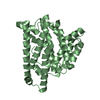
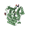
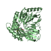
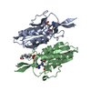
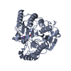
| Title | Crystal Structure of the Putative Selenoprotein W-related family Protein from Agrobacterium tumefaciens | ||||||
 Components Components | hypothetical protein Atu0228 | ||||||
 Keywords Keywords | STRUCTURAL GENOMICS / UNKNOWN FUNCTION / alph-beta structure / 4 helix bundle / PSI / Protein Structure Initiative / Midwest Center for Structural Genomics / MCSG | ||||||
| Function / homology | Selenoprotein, Rdx-type / Rdx family / Glutaredoxin / Glutaredoxin / Thioredoxin-like superfamily / 3-Layer(aba) Sandwich / Alpha Beta / : / SelT/selW/selH selenoprotein Function and homology information Function and homology information | ||||||
| Biological species |  Agrobacterium tumefaciens str. (bacteria) Agrobacterium tumefaciens str. (bacteria) | ||||||
| Method |  X-RAY DIFFRACTION / SYNCHROTRON / MAD / Resolution: 1.9 Å X-RAY DIFFRACTION / SYNCHROTRON / MAD / Resolution: 1.9 Å | ||||||
 Authors Authors | Kim, Y. / Joachimiak, A. / Xu, X. / Zheng, H. / Savchenko, A. / Edwards, A. / Midwest Center for Structural Genomics (MCSG) | ||||||
 Citation Citation | Journal: To be Published / Year: 2006 Title: Crystal Structure of the Putative Selenoprotein W-related family Protein from Agrobacterium tumefaciens Authors: Kim, Y. / Xu, X. / Zheng, H. / Savchenko, A. / Edwards, A. / Joachimiak, A. | ||||||
| History |
|
- Structure visualization
| Structure viewer | Molecule: MolmilJmol/JSmol |
|---|
- Downloads & links
-Download
| PDBx/mmCIF format | 2fa8.cif.gz | 85.4 KB | Display | PDBx/mmCIF format |
|---|---|---|---|---|
| PDB format | pdb2fa8.ent.gz | 65.3 KB | Display | PDB format |
| PDBx/mmJSON format | 2fa8.json.gz | Tree view | PDBx/mmJSON format | |
| Others |  Other downloads Other downloads |
-Validation report
| Arichive directory | https://data.pdbj.org/pub/pdb/validation_reports/fa/2fa8ftp://data.pdbj.org/pub/pdb/validation_reports/fa/2fa8 | HTTPS FTP |
|---|
-Related structure data
| Similar structure data | |
|---|---|
| Other databases |


-Links
 PDBj
PDBj- Assembly
| Deposited unit | 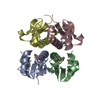
| ||||||||
|---|---|---|---|---|---|---|---|---|---|
| 1 |
| ||||||||
| Unit cell |
|
-Components
| #1: Protein | Mass: 11959.262 Da / Num. of mol.: 4 Source method: isolated from a genetically manipulated source Source: (gene. exp.) Agrobacterium tumefaciens str. (bacteria)Species: Agrobacterium tumefaciens / Strain: C58 / Gene: atu0228 / Plasmid: pMCSG7 / Species (production host): Escherichia coli / Production host: #2: Water | ChemComp-HOH / |  Mass: 18.015 Da / Num. of mol.: 198 / Source method: isolated from a natural source / Formula: H2O Mass: 18.015 Da / Num. of mol.: 198 / Source method: isolated from a natural source / Formula: H2OHas protein modification | Y | |
|---|
-Experimental details
-Experiment
| Experiment | Method: X-RAY DIFFRACTION / Number of used crystals: 2 |
|---|
- Sample preparation
| Crystal | Density Matthews: 1.84 Å3/Da / Density % sol: 33.03 % |
|---|---|
| Crystal grow | Temperature: 295 K / Method: vapor diffusion, hanging drop / pH: 7.5 Details: 0.2M potassium chloride, 20% PEG3350, pH 7.5, VAPOR DIFFUSION, HANGING DROP, temperature 295K |
-Data collection
| Diffraction |
| ||||||||||||||||||
|---|---|---|---|---|---|---|---|---|---|---|---|---|---|---|---|---|---|---|---|
| Diffraction source |
| ||||||||||||||||||
| Detector |
| ||||||||||||||||||
| Radiation |
| ||||||||||||||||||
| Radiation wavelength |
| ||||||||||||||||||
| Reflection | Resolution: 1.9→46.42 Å / Num. all: 27903 / Num. obs: 29741 / % possible obs: 97.9 % / Observed criterion σ(F): 0 / Observed criterion σ(I): 0 / Redundancy: 6.6 % / Rmerge(I) obs: 0.085 / Net I/σ(I): 8.7 | ||||||||||||||||||
| Reflection shell | Resolution: 1.9→1.97 Å / Redundancy: 4.3 % / Rmerge(I) obs: 0.496 / Mean I/σ(I) obs: 1.9 / Num. unique all: 2341 / % possible all: 84.3 |

- Processing
| Software |
| ||||||||||||||||||||||||||||||||||||||||||||||||||||||||||||||||||||||||||||||||||||||||||||||||||||||||||||||||||||||||||||||||||||||||||||||||||||||||||||||||||||||||||
|---|---|---|---|---|---|---|---|---|---|---|---|---|---|---|---|---|---|---|---|---|---|---|---|---|---|---|---|---|---|---|---|---|---|---|---|---|---|---|---|---|---|---|---|---|---|---|---|---|---|---|---|---|---|---|---|---|---|---|---|---|---|---|---|---|---|---|---|---|---|---|---|---|---|---|---|---|---|---|---|---|---|---|---|---|---|---|---|---|---|---|---|---|---|---|---|---|---|---|---|---|---|---|---|---|---|---|---|---|---|---|---|---|---|---|---|---|---|---|---|---|---|---|---|---|---|---|---|---|---|---|---|---|---|---|---|---|---|---|---|---|---|---|---|---|---|---|---|---|---|---|---|---|---|---|---|---|---|---|---|---|---|---|---|---|---|---|---|---|---|---|---|
| Refinement | Method to determine structure: MAD / Resolution: 1.9→46.42 Å / Cor.coef. Fo:Fc: 0.957 / Cor.coef. Fo:Fc free: 0.931 / SU B: 4.019 / SU ML: 0.119 / Cross valid method: THROUGHOUT / σ(F): 0 / ESU R: 0.173 / ESU R Free: 0.161 / Stereochemistry target values: MAXIMUM LIKELIHOOD
| ||||||||||||||||||||||||||||||||||||||||||||||||||||||||||||||||||||||||||||||||||||||||||||||||||||||||||||||||||||||||||||||||||||||||||||||||||||||||||||||||||||||||||
| Solvent computation | Ion probe radii: 0.8 Å / Shrinkage radii: 0.8 Å / VDW probe radii: 1.2 Å / Solvent model: MASK | ||||||||||||||||||||||||||||||||||||||||||||||||||||||||||||||||||||||||||||||||||||||||||||||||||||||||||||||||||||||||||||||||||||||||||||||||||||||||||||||||||||||||||
| Displacement parameters | Biso mean: 31.986 Å2
| ||||||||||||||||||||||||||||||||||||||||||||||||||||||||||||||||||||||||||||||||||||||||||||||||||||||||||||||||||||||||||||||||||||||||||||||||||||||||||||||||||||||||||
| Refinement step | Cycle: LAST / Resolution: 1.9→46.42 Å
| ||||||||||||||||||||||||||||||||||||||||||||||||||||||||||||||||||||||||||||||||||||||||||||||||||||||||||||||||||||||||||||||||||||||||||||||||||||||||||||||||||||||||||
| Refine LS restraints |
| ||||||||||||||||||||||||||||||||||||||||||||||||||||||||||||||||||||||||||||||||||||||||||||||||||||||||||||||||||||||||||||||||||||||||||||||||||||||||||||||||||||||||||
| LS refinement shell | Resolution: 1.9→1.949 Å / Total num. of bins used: 20
|
