 Movie
Movie Controller

[English] 日本語
 Yorodumi
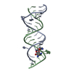
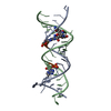
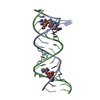
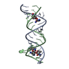
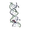
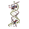
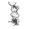
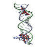
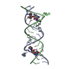
- PDB-2o3x: Crystal Structure of the Prokaryotic Ribosomal Decoding Site Comp... -
+ Open data

- Basic information
| Entry | Database: PDB / ID: 2o3x | ||||||||||||||||||
|---|---|---|---|---|---|---|---|---|---|---|---|---|---|---|---|---|---|---|---|
| Title | Crystal Structure of the Prokaryotic Ribosomal Decoding Site Complexed with Paromamine Derivative NB30 | ||||||||||||||||||
 Components Components | RNA (5'-R(* Keywords KeywordsRNA / aminoglycoside / antibiotics / ribosome / decoding site / Prokaryote / Translation inhibition / Stop codon readthrough | Function / homology | Chem-N30 / RNA / RNA (> 10) |  Function and homology information Function and homology informationMethod |  X-RAY DIFFRACTION / SYNCHROTRON / MOLECULAR REPLACEMENT / Resolution: 2.9 Å X-RAY DIFFRACTION / SYNCHROTRON / MOLECULAR REPLACEMENT / Resolution: 2.9 Å  Authors AuthorsKondo, J. / Hainrichson, M. / Nudelman, I. / Shallom-Shezifi, D. / Baasov, T. / Westhof, E. |  CitationJournal: Chembiochem / Year: 2007 CitationJournal: Chembiochem / Year: 2007Title: Differential Selectivity of Natural and Synthetic Aminoglycosides towards the Eukaryotic and Prokaryotic Decoding A Sites. Authors: Kondo, J. / Hainrichson, M. / Nudelman, I. / Shallom-Shezifi, D. / Barbieri, C.M. / Pilch, D.S. / Westhof, E. / Baasov, T. History |
|
- Structure visualization
| Structure viewer | Molecule: MolmilJmol/JSmol |
|---|
- Downloads & links
-Download
| PDBx/mmCIF format | 2o3x.cif.gz | 34.5 KB | Display | PDBx/mmCIF format |
|---|---|---|---|---|
| PDB format | pdb2o3x.ent.gz | 23.1 KB | Display | PDB format |
| PDBx/mmJSON format | 2o3x.json.gz | Tree view | PDBx/mmJSON format | |
| Others |  Other downloads Other downloads |
-Validation report
| Arichive directory | https://data.pdbj.org/pub/pdb/validation_reports/o3/2o3xftp://data.pdbj.org/pub/pdb/validation_reports/o3/2o3x | HTTPS FTP |
|---|
-Related structure data
| Related structure data |  2o3vC  2o3wC  2o3yC 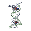 1j7tS C: citing same article ( S: Starting model for refinement |
|---|---|
| Similar structure data |

-Links
 PDBj
PDBj


- Assembly
| Deposited unit | 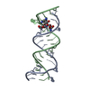
| ||||||||
|---|---|---|---|---|---|---|---|---|---|
| 1 |
| ||||||||
| Unit cell |
|
-Components
| #1: RNA chain | Mass: 7355.409 Da / Num. of mol.: 2 / Source method: obtained synthetically / Details: Chemically synthesized #2: Chemical | ChemComp-N30 / ( |   Mass: 454.473 Da / Num. of mol.: 1 / Source method: obtained synthetically / Formula: C17H34N4O10 Mass: 454.473 Da / Num. of mol.: 1 / Source method: obtained synthetically / Formula: C17H34N4O10 |
|---|
-Experimental details
-Experiment
| Experiment | Method: X-RAY DIFFRACTION / Number of used crystals: 1 |
|---|
- Sample preparation
| Crystal | Density Matthews: 2.26 Å3/Da / Density % sol: 45.59 % | ||||||||||||||||||||||||||||||||||||||||
|---|---|---|---|---|---|---|---|---|---|---|---|---|---|---|---|---|---|---|---|---|---|---|---|---|---|---|---|---|---|---|---|---|---|---|---|---|---|---|---|---|---|
| Crystal grow | Temperature: 300 K / Method: vapor diffusion, hanging drop / pH: 6.5 Details: Sodium Cacodylate, Potassium chloride, 2-methyl-2,4-pentanediol, spermine tetrahydrochloride, glycerol, pH 6.5, VAPOR DIFFUSION, HANGING DROP, temperature 300K | ||||||||||||||||||||||||||||||||||||||||
| Components of the solutions |
|
-Data collection
| Diffraction | Mean temperature: 100 K | ||||||||||||||||||||||||||||||||||||||||||||||||||||||||||||||||||||||||||||||||||||||||
|---|---|---|---|---|---|---|---|---|---|---|---|---|---|---|---|---|---|---|---|---|---|---|---|---|---|---|---|---|---|---|---|---|---|---|---|---|---|---|---|---|---|---|---|---|---|---|---|---|---|---|---|---|---|---|---|---|---|---|---|---|---|---|---|---|---|---|---|---|---|---|---|---|---|---|---|---|---|---|---|---|---|---|---|---|---|---|---|---|---|
| Diffraction source | Source: SYNCHROTRON / Site: ESRF  / Beamline: ID29 / Wavelength: 0.9737 Å / Beamline: ID29 / Wavelength: 0.9737 Å | ||||||||||||||||||||||||||||||||||||||||||||||||||||||||||||||||||||||||||||||||||||||||
| Detector | Type: ADSC QUANTUM 315 / Detector: CCD / Date: May 22, 2006 | ||||||||||||||||||||||||||||||||||||||||||||||||||||||||||||||||||||||||||||||||||||||||
| Radiation | Monochromator: Si(111) / Protocol: SINGLE WAVELENGTH / Monochromatic (M) / Laue (L): M / Scattering type: x-ray | ||||||||||||||||||||||||||||||||||||||||||||||||||||||||||||||||||||||||||||||||||||||||
| Radiation wavelength | Wavelength: 0.9737 Å / Relative weight: 1 | ||||||||||||||||||||||||||||||||||||||||||||||||||||||||||||||||||||||||||||||||||||||||
| Reflection | Resolution: 2.9→42.86 Å / Num. obs: 3222 / % possible obs: 99.4 % / Redundancy: 6.53 % / Rmerge(I) obs: 0.163 / Χ2: 0.94 / Net I/σ(I): 8.3 / Scaling rejects: 159 | ||||||||||||||||||||||||||||||||||||||||||||||||||||||||||||||||||||||||||||||||||||||||
| Reflection shell |
|
- Processing
| Software |
| ||||||||||||||||||||||||||||||||||||||||||||||||||||||
|---|---|---|---|---|---|---|---|---|---|---|---|---|---|---|---|---|---|---|---|---|---|---|---|---|---|---|---|---|---|---|---|---|---|---|---|---|---|---|---|---|---|---|---|---|---|---|---|---|---|---|---|---|---|---|---|
| Refinement | Method to determine structure: MOLECULAR REPLACEMENT Starting model: 1J7T Resolution: 2.9→10 Å / FOM work R set: 0.756 Isotropic thermal model: G. Parkinson, J. Vojtechovsky, L. Clowney, A.T. Brunger, H.M. Berman, New Parameters for the Refinement of Nucleic Acid Containing Structures, Acta Cryst. D, 52, 57-64 (1996). σ(F): 3
| ||||||||||||||||||||||||||||||||||||||||||||||||||||||
| Solvent computation | Bsol: 47.316 Å2 | ||||||||||||||||||||||||||||||||||||||||||||||||||||||
| Displacement parameters | Biso mean: 76.921 Å2
| ||||||||||||||||||||||||||||||||||||||||||||||||||||||
| Refinement step | Cycle: LAST / Resolution: 2.9→10 Å
| ||||||||||||||||||||||||||||||||||||||||||||||||||||||
| Refine LS restraints |
| ||||||||||||||||||||||||||||||||||||||||||||||||||||||
| LS refinement shell | Refine-ID: X-RAY DIFFRACTION / Total num. of bins used: 8
| ||||||||||||||||||||||||||||||||||||||||||||||||||||||
| Xplor file |
|
