 Movie
Movie Controller

[English] 日本語
 Yorodumi
- PDB-4p20: Crystal structures of the bacterial ribosomal decoding site compl... -
+ Open data

- Basic information
| Entry | Database: PDB / ID: 4p20 | |||||||||||||||||||||||
|---|---|---|---|---|---|---|---|---|---|---|---|---|---|---|---|---|---|---|---|---|---|---|---|---|
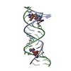
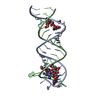
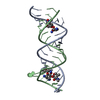
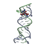
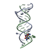
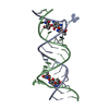
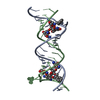
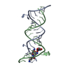
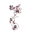
| Title | Crystal structures of the bacterial ribosomal decoding site complexed with amikacin | |||||||||||||||||||||||
 Components Components | 5'-R(* Keywords KeywordsRNA/ANTIBIOTIC / AMINOGLYCOSIDE / HABA GROUP / RIBOSOMAL DECODING SITE / X-RAY ANALYSIS / RNA / AMIKACIN / RNA-ANTIBIOTIC complex | Function / homology | Chem-AKN / RNA / RNA (> 10) |  Function and homology information Function and homology informationBiological species | synthetic construct (others) | Method |  X-RAY DIFFRACTION / SYNCHROTRON / MOLECULAR REPLACEMENT / molecular replacement / Resolution: 2.7 Å X-RAY DIFFRACTION / SYNCHROTRON / MOLECULAR REPLACEMENT / molecular replacement / Resolution: 2.7 Å  Authors AuthorsKondo, J. / Francois, B. / Russell, R.J.M. / Murray, J.B. / Westhof, E. |  CitationJournal: Biochimie / Year: 2006 CitationJournal: Biochimie / Year: 2006Title: Crystal structure of the bacterial ribosomal decoding site complexed with amikacin containing the gamma-amino-alpha-hydroxybutyryl (haba) group. Authors: Kondo, J. / Francois, B. / Russell, R.J. / Murray, J.B. / Westhof, E. History |
|
- Structure visualization
| Structure viewer | Molecule: MolmilJmol/JSmol |
|---|
- Downloads & links
-Download
| PDBx/mmCIF format | 4p20.cif.gz | 38.8 KB | Display | PDBx/mmCIF format |
|---|---|---|---|---|
| PDB format | pdb4p20.ent.gz | 26 KB | Display | PDB format |
| PDBx/mmJSON format | 4p20.json.gz | Tree view | PDBx/mmJSON format | |
| Others |  Other downloads Other downloads |
-Validation report
| Arichive directory | https://data.pdbj.org/pub/pdb/validation_reports/p2/4p20ftp://data.pdbj.org/pub/pdb/validation_reports/p2/4p20 | HTTPS FTP |
|---|
-Related structure data
| Similar structure data |
|---|

-Links
 PDBj
PDBj


- Assembly
| Deposited unit | 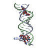
| ||||||||
|---|---|---|---|---|---|---|---|---|---|
| 1 |
| ||||||||
| Unit cell |
|
-Components
| #1: RNA chain | Mass: 7355.409 Da / Num. of mol.: 2 / Source method: obtained synthetically / Source: (synth.) synthetic construct (others) #2: Chemical |   Mass: 585.603 Da / Num. of mol.: 2 / Source method: obtained synthetically / Formula: C22H43N5O13 Mass: 585.603 Da / Num. of mol.: 2 / Source method: obtained synthetically / Formula: C22H43N5O13#3: Water | ChemComp-HOH / |  Mass: 18.015 Da / Num. of mol.: 75 / Source method: isolated from a natural source / Formula: H2O Mass: 18.015 Da / Num. of mol.: 75 / Source method: isolated from a natural source / Formula: H2O |
|---|
-Experimental details
-Experiment
| Experiment | Method: X-RAY DIFFRACTION / Number of used crystals: 1 |
|---|
- Sample preparation
| Crystal | Density Matthews: 2.65 Å3/Da / Density % sol: 53.51 % |
|---|---|
| Crystal grow | Temperature: 310 K / Method: vapor diffusion / pH: 6.4 / Details: MPF, Na Cacodylate, NaCl, KCl |
-Data collection
| Diffraction | Mean temperature: 100 K | |||||||||||||||||||||||||||||||||||||||||||||||||||||||||||||||||||||||||||||||||||||||||||||||||||
|---|---|---|---|---|---|---|---|---|---|---|---|---|---|---|---|---|---|---|---|---|---|---|---|---|---|---|---|---|---|---|---|---|---|---|---|---|---|---|---|---|---|---|---|---|---|---|---|---|---|---|---|---|---|---|---|---|---|---|---|---|---|---|---|---|---|---|---|---|---|---|---|---|---|---|---|---|---|---|---|---|---|---|---|---|---|---|---|---|---|---|---|---|---|---|---|---|---|---|---|---|
| Diffraction source | Source: SYNCHROTRON / Site: ESRF  / Beamline: ID14-4 / Wavelength: 0.9357 Å / Beamline: ID14-4 / Wavelength: 0.9357 Å | |||||||||||||||||||||||||||||||||||||||||||||||||||||||||||||||||||||||||||||||||||||||||||||||||||
| Detector | Type: ADSC QUANTUM 4 / Detector: CCD / Date: Jun 26, 2004 | |||||||||||||||||||||||||||||||||||||||||||||||||||||||||||||||||||||||||||||||||||||||||||||||||||
| Radiation | Protocol: SINGLE WAVELENGTH / Scattering type: x-ray | |||||||||||||||||||||||||||||||||||||||||||||||||||||||||||||||||||||||||||||||||||||||||||||||||||
| Radiation wavelength | Wavelength: 0.9357 Å / Relative weight: 1 | |||||||||||||||||||||||||||||||||||||||||||||||||||||||||||||||||||||||||||||||||||||||||||||||||||
| Reflection | Resolution: 2.7→32.82 Å / Num. obs: 4398 / % possible obs: 100 % / Redundancy: 3.59 % / Rmerge(I) obs: 0.16 / Χ2: 0.96 / Net I/σ(I): 4.8 / Num. measured all: 15897 / Scaling rejects: 120 | |||||||||||||||||||||||||||||||||||||||||||||||||||||||||||||||||||||||||||||||||||||||||||||||||||
| Reflection shell | Diffraction-ID: 1
|
-Phasing
| Phasing | Method: molecular replacement |
|---|
- Processing
| Software |
| ||||||||||||||||||||||||
|---|---|---|---|---|---|---|---|---|---|---|---|---|---|---|---|---|---|---|---|---|---|---|---|---|---|
| Refinement | Method to determine structure: MOLECULAR REPLACEMENT / Resolution: 2.7→10 Å / FOM work R set: 0.807 / Cross valid method: FREE R-VALUE / σ(F): 179
| ||||||||||||||||||||||||
| Solvent computation | Bsol: 64.5977 Å2 | ||||||||||||||||||||||||
| Displacement parameters | Biso max: 120.73 Å2 / Biso mean: 45.5528 Å2 / Biso min: 10.2 Å2
| ||||||||||||||||||||||||
| Refinement step | Cycle: final / Resolution: 2.7→10 Å
| ||||||||||||||||||||||||
| Refine LS restraints |
| ||||||||||||||||||||||||
| Xplor file |
|
