 Movie
Movie Controller

[English] 日本語
 Yorodumi
- PDB-2o0f: Docking of the modified RF3 X-ray structure into cryo-EM map of E... -
+ Open data

- Basic information
| Entry | Database: PDB / ID: 2o0f | ||||||
|---|---|---|---|---|---|---|---|
| Title | Docking of the modified RF3 X-ray structure into cryo-EM map of E.coli 70S ribosome bound with RF3 | ||||||
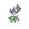
 Components Components | Peptide chain release factor 3 | ||||||
 Keywords Keywords | TRANSLATION / RF3 / Ribosome / cryo-EM | ||||||
| Function / homology |  Function and homology information Function and homology informationregulation of translational termination / translation release factor activity, codon specific / translation release factor activity, codon nonspecific / guanosine tetraphosphate binding / translational termination / maintenance of translational fidelity / GDP binding / GTPase activity / GTP binding / cytoplasm / cytosol Similarity search - Function | ||||||
| Biological species |  | ||||||
| Method | ELECTRON MICROSCOPY / single particle reconstruction / cryo EM / Resolution: 15.5 Å | ||||||
 Authors Authors | Gao, H. / Zhou, Z. / Rawat, U. / Huang, C. / Bouakaz, L. / Wang, C. / Liu, Y. / Zavialov, A. / Gursky, R. / Sanyal, S. ...Gao, H. / Zhou, Z. / Rawat, U. / Huang, C. / Bouakaz, L. / Wang, C. / Liu, Y. / Zavialov, A. / Gursky, R. / Sanyal, S. / Ehrenberg, M. / Frank, J. / Song, H. | ||||||
 Citation Citation | Journal: Cell / Year: 2007 Title: RF3 induces ribosomal conformational changes responsible for dissociation of class I release factors. Authors: Haixiao Gao / Zhihong Zhou / Urmila Rawat / Chenhui Huang / Lamine Bouakaz / Chernhoe Wang / Zhihong Cheng / Yuying Liu / Andrey Zavialov / Richard Gursky / Suparna Sanyal / Måns Ehrenberg ...Authors: Haixiao Gao / Zhihong Zhou / Urmila Rawat / Chenhui Huang / Lamine Bouakaz / Chernhoe Wang / Zhihong Cheng / Yuying Liu / Andrey Zavialov / Richard Gursky / Suparna Sanyal / Måns Ehrenberg / Joachim Frank / Haiwei Song /  Abstract: During translation termination, class II release factor RF3 binds to the ribosome to promote rapid dissociation of a class I release factor (RF) in a GTP-dependent manner. We present the crystal ...During translation termination, class II release factor RF3 binds to the ribosome to promote rapid dissociation of a class I release factor (RF) in a GTP-dependent manner. We present the crystal structure of E. coli RF3*GDP, which has a three-domain architecture strikingly similar to the structure of EF-Tu*GTP. Biochemical data on RF3 mutants show that a surface region involving domains II and III is important for distinct steps in the action cycle of RF3. Furthermore, we present a cryo-electron microscopy (cryo-EM) structure of the posttermination ribosome bound with RF3 in the GTP form. Our data show that RF3*GTP binding induces large conformational changes in the ribosome, which break the interactions of the class I RF with both the decoding center and the GTPase-associated center of the ribosome, apparently leading to the release of the class I RF. | ||||||
| History |
|
- Structure visualization
| Movie |
Movie viewer |
|---|---|
| Structure viewer | Molecule: MolmilJmol/JSmol |
- Downloads & links
-Download
| PDBx/mmCIF format | 2o0f.cif.gz | 28.1 KB | Display | PDBx/mmCIF format |
|---|---|---|---|---|
| PDB format | pdb2o0f.ent.gz | 12.7 KB | Display | PDB format |
| PDBx/mmJSON format | 2o0f.json.gz | Tree view | PDBx/mmJSON format | |
| Others |  Other downloads Other downloads |
-Validation report
| Arichive directory | https://data.pdbj.org/pub/pdb/validation_reports/o0/2o0fftp://data.pdbj.org/pub/pdb/validation_reports/o0/2o0f | HTTPS FTP |
|---|
-Related structure data
| Related structure data | 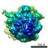 1302MC  2h5eC C: citing same article ( M: map data used to model this data |
|---|---|
| Similar structure data |


-Links
 PDBj
PDBj


- Assembly
| Deposited unit | 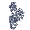
|
|---|---|
| 1 |
|
-Components
| #1: Protein | Mass: 59651.934 Da / Num. of mol.: 1 Source method: isolated from a genetically manipulated source Source: (gene. exp.) |
|---|
-Experimental details
-Experiment
| Experiment | Method: ELECTRON MICROSCOPY |
|---|---|
| EM experiment | Aggregation state: PARTICLE / 3D reconstruction method: single particle reconstruction |
- Sample preparation
| Component | Name: ribosomal release complex bound with RF3 / Type: RIBOSOME |
|---|---|
| Buffer solution | Name: Polymix Buffer / pH: 7.5 / Details: Polymix Buffer |
| Specimen | Conc.: 0.08 mg/ml / Embedding applied: NO / Shadowing applied: NO / Staining applied: NO / Vitrification applied: YES |
| Specimen support | Details: Quanti-foil grids coated with a thin carbon layer |
| Vitrification | Instrument: FEI VITROBOT MARK I / Cryogen name: ETHANE / Details: Rapid-freezing in liquid ethane |
- Electron microscopy imaging
| Experimental equipment |  Model: Tecnai F20 / Image courtesy: FEI Company |
|---|---|
| Microscopy | Model: FEI TECNAI F20 / Date: Jan 1, 2002 |
| Electron gun | Electron source:  FIELD EMISSION GUN / Accelerating voltage: 200 kV / Illumination mode: FLOOD BEAM FIELD EMISSION GUN / Accelerating voltage: 200 kV / Illumination mode: FLOOD BEAM |
| Electron lens | Mode: BRIGHT FIELD / Nominal magnification: 50000 X / Calibrated magnification: 49696 X / Nominal defocus max: 4000 nm / Nominal defocus min: 2000 nm / Cs: 2 mm |
| Specimen holder | Temperature: 93 K / Tilt angle max: 0 ° / Tilt angle min: 0 ° |
| Image recording | Electron dose: 20 e/Å2 / Film or detector model: KODAK SO-163 FILM |
| Radiation | Protocol: SINGLE WAVELENGTH / Monochromatic (M) / Laue (L): M / Scattering type: x-ray |
| Radiation wavelength | Relative weight: 1 |
- Processing
| EM software |
| ||||||||||||||||
|---|---|---|---|---|---|---|---|---|---|---|---|---|---|---|---|---|---|
| CTF correction | Details: CTF correction of 3D map | ||||||||||||||||
| Symmetry | Point symmetry: C1 (asymmetric) | ||||||||||||||||
| 3D reconstruction | Method: Single particle, Reference-based alignment / Resolution: 15.5 Å / Resolution method: FSC 0.5 CUT-OFF / Num. of particles: 45000 / Nominal pixel size: 2.8 Å / Actual pixel size: 2.82 Å / Magnification calibration: TMV Details: SPIDER package, 0.5 cutoff of FSC, This entry contains CA atoms only Symmetry type: POINT | ||||||||||||||||
| Atomic model building | Protocol: RIGID BODY FIT / Space: REAL / Target criteria: cross-correlation coefficient Details: METHOD--auto REFINEMENT PROTOCOL--Multi-rigid body, real-space refinement | ||||||||||||||||
| Atomic model building | PDB-ID: 2H5E Accession code: 2H5E / Source name: PDB / Type: experimental model | ||||||||||||||||
| Refinement step | Cycle: LAST
|
