 Movie
Movie Controller

[English] 日本語
 Yorodumi
- PDB-2nyl: Crystal structure of Protein Phosphatase 2A (PP2A) holoenzyme wit... -
+ Open data

- Basic information
| Entry | Database: PDB / ID: 2nyl | |||||||||
|---|---|---|---|---|---|---|---|---|---|---|
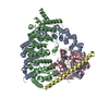
| Title | Crystal structure of Protein Phosphatase 2A (PP2A) holoenzyme with the catalytic subunit carboxyl terminus truncated | |||||||||
 Components Components |
| |||||||||
 Keywords Keywords | HYDROLASE/HYDROLASE INHIBITOR / HEAT repeat / HYDROLASE-HYDROLASE INHIBITOR COMPLEX | |||||||||
| Function / homology |  Function and homology information Function and homology informationmeiotic spindle elongation / PP2A-mediated dephosphorylation of key metabolic factors / RNA polymerase II CTD heptapeptide repeat S2 phosphatase activity / RNA polymerase II CTD heptapeptide repeat S7 phosphatase activity / peptidyl-threonine dephosphorylation / regulation of meiotic cell cycle process involved in oocyte maturation / mitotic sister chromatid separation / MASTL Facilitates Mitotic Progression / protein phosphatase type 2A complex / meiotic sister chromatid cohesion, centromeric ...meiotic spindle elongation / PP2A-mediated dephosphorylation of key metabolic factors / RNA polymerase II CTD heptapeptide repeat S2 phosphatase activity / RNA polymerase II CTD heptapeptide repeat S7 phosphatase activity / peptidyl-threonine dephosphorylation / regulation of meiotic cell cycle process involved in oocyte maturation / mitotic sister chromatid separation / MASTL Facilitates Mitotic Progression / protein phosphatase type 2A complex / meiotic sister chromatid cohesion, centromeric / INTAC complex / RNA polymerase II CTD heptapeptide repeat S5 phosphatase activity / FAR/SIN/STRIPAK complex / female meiotic nuclear division / Regulation of glycolysis by fructose 2,6-bisphosphate metabolism / meiotic sister chromatid cohesion / Inhibition of replication initiation of damaged DNA by RB1/E2F1 / protein phosphatase regulator activity / protein antigen binding / GABA receptor binding / APC truncation mutants have impaired AXIN binding / AXIN missense mutants destabilize the destruction complex / Truncations of AMER1 destabilize the destruction complex / positive regulation of extrinsic apoptotic signaling pathway in absence of ligand / ERKs are inactivated / Initiation of Nuclear Envelope (NE) Reformation / Beta-catenin phosphorylation cascade / Signaling by GSK3beta mutants / CTNNB1 S33 mutants aren't phosphorylated / CTNNB1 S37 mutants aren't phosphorylated / CTNNB1 S45 mutants aren't phosphorylated / CTNNB1 T41 mutants aren't phosphorylated / Co-stimulation by CD28 / RNA polymerase II transcription initiation surveillance / regulation of growth / Disassembly of the destruction complex and recruitment of AXIN to the membrane / protein dephosphorylation / negative regulation of epithelial to mesenchymal transition / Co-inhibition by CTLA4 / Platelet sensitization by LDL / protein-serine/threonine phosphatase / negative regulation of glycolytic process through fructose-6-phosphate / ERK/MAPK targets / vascular endothelial cell response to oscillatory fluid shear stress / protein serine/threonine phosphatase activity / T cell homeostasis / mesoderm development / positive regulation of NLRP3 inflammasome complex assembly / regulation of cell differentiation / regulation of microtubule polymerization / regulation of G1/S transition of mitotic cell cycle / protein phosphatase activator activity / lateral plasma membrane / intrinsic apoptotic signaling pathway in response to DNA damage by p53 class mediator / chromosome, centromeric region / DARPP-32 events / negative regulation of hippo signaling / Cyclin A/B1/B2 associated events during G2/M transition / Nonsense Mediated Decay (NMD) enhanced by the Exon Junction Complex (EJC) / spindle assembly / phosphoprotein phosphatase activity / Loss of Nlp from mitotic centrosomes / Loss of proteins required for interphase microtubule organization from the centrosome / Amplification of signal from unattached kinetochores via a MAD2 inhibitory signal / Recruitment of mitotic centrosome proteins and complexes / protein tyrosine phosphatase activity / Recruitment of NuMA to mitotic centrosomes / Anchoring of the basal body to the plasma membrane / Mitotic Prometaphase / EML4 and NUDC in mitotic spindle formation / Turbulent (oscillatory, disturbed) flow shear stress activates signaling by PIEZO1 and integrins in endothelial cells / AURKA Activation by TPX2 / Resolution of Sister Chromatid Cohesion / negative regulation of phosphatidylinositol 3-kinase/protein kinase B signal transduction / meiotic cell cycle / DNA damage response, signal transduction by p53 class mediator / chromosome segregation / negative regulation of canonical Wnt signaling pathway / RAF activation / RHO GTPases Activate Formins / Spry regulation of FGF signaling / PKR-mediated signaling / response to lead ion / Degradation of beta-catenin by the destruction complex / tau protein binding / spindle pole / Cyclin D associated events in G1 / Negative regulation of MAPK pathway / Separation of Sister Chromatids / Regulation of TP53 Degradation / Regulation of PLK1 Activity at G2/M Transition / mitotic cell cycle / PI5P, PP2A and IER3 Regulate PI3K/AKT Signaling / microtubule cytoskeleton / protein-containing complex assembly / proteasome-mediated ubiquitin-dependent protein catabolic process / neuron projection / intracellular signal transduction / membrane raft / protein heterodimerization activity Similarity search - Function | |||||||||
| Biological species |  Homo sapiens (human) Homo sapiens (human) Cyanobacteria (cyanobacteria) Cyanobacteria (cyanobacteria) | |||||||||
| Method |  X-RAY DIFFRACTION / SYNCHROTRON / MOLECULAR REPLACEMENT / Resolution: 3.8 Å X-RAY DIFFRACTION / SYNCHROTRON / MOLECULAR REPLACEMENT / Resolution: 3.8 Å | |||||||||
 Authors Authors | Xing, Y. / Xu, Y. / Chen, Y. / Chao, Y. / Lin, Z. / Shi, Y. | |||||||||
 Citation Citation | Journal: Cell(Cambridge,Mass.) / Year: 2006 Title: Structure of the Protein Phosphatase 2A Holoenzyme. Authors: Xu, Y. / Xing, Y. / Chen, Y. / Chao, Y. / Lin, Z. / Fan, E. / Yu, J.W. / Strack, S. / Jeffrey, P.D. / Shi, Y. | |||||||||
| History |
|
- Structure visualization
| Structure viewer | Molecule: MolmilJmol/JSmol |
|---|
- Downloads & links
-Download
| PDBx/mmCIF format | 2nyl.cif.gz | 517.3 KB | Display | PDBx/mmCIF format |
|---|---|---|---|---|
| PDB format | pdb2nyl.ent.gz | 415.8 KB | Display | PDB format |
| PDBx/mmJSON format | 2nyl.json.gz | Tree view | PDBx/mmJSON format | |
| Others |  Other downloads Other downloads |
-Validation report
| Arichive directory | https://data.pdbj.org/pub/pdb/validation_reports/ny/2nylftp://data.pdbj.org/pub/pdb/validation_reports/ny/2nyl | HTTPS FTP |
|---|
-Related structure data
| Related structure data |  2nppSC  2nymC S: Starting model for refinement C: citing same article ( |
|---|---|
| Similar structure data |


-Links
 PDBj
PDBj


- Assembly
| Deposited unit | 
| ||||||||
|---|---|---|---|---|---|---|---|---|---|
| 1 | 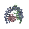
| ||||||||
| 2 | 
| ||||||||
| Unit cell |
| ||||||||
| Details | Each holoenzyme contains one copy of catalytic subunit, one copy of scaffolding subunit and one copy of the regulatory subunit. |
-Components
| #1: Protein | Mass: 65356.316 Da / Num. of mol.: 2 Source method: isolated from a genetically manipulated source Source: (gene. exp.) Homo sapiens (human) / Gene: PPP2R1A / Plasmid: pGEX-2T / Species (production host): Escherichia coli / Production host: #2: Protein | Mass: 46467.258 Da / Num. of mol.: 2 Source method: isolated from a genetically manipulated source Source: (gene. exp.) Homo sapiens (human) / Gene: PPP2R5C, KIAA0044 / Plasmid: pGEX-2T / Species (production host): Escherichia coli / Production host: #3: Protein | Mass: 33674.910 Da / Num. of mol.: 2 Source method: isolated from a genetically manipulated source Source: (gene. exp.) Homo sapiens (human) / Gene: PPP2CA / Production host:   Spodoptera frugiperda (fall armyworm) / Strain (production host): Hi-5 Spodoptera frugiperda (fall armyworm) / Strain (production host): Hi-5References: UniProt: P67775, protein-serine/threonine phosphatase #4: Protein/peptide | 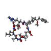  Type: Oligopeptide / Class: Toxin / Mass: 1014.195 Da / Num. of mol.: 2 / Source method: obtained synthetically / Source: (synth.) Cyanobacteria (cyanobacteria) / References: NOR: NOR00109, Microcystin LR Type: Oligopeptide / Class: Toxin / Mass: 1014.195 Da / Num. of mol.: 2 / Source method: obtained synthetically / Source: (synth.) Cyanobacteria (cyanobacteria) / References: NOR: NOR00109, Microcystin LR#5: Chemical | ChemComp-MN /   Mass: 54.938 Da / Num. of mol.: 4 / Source method: obtained synthetically / Formula: Mn Mass: 54.938 Da / Num. of mol.: 4 / Source method: obtained synthetically / Formula: Mn |
|---|
-Experimental details
-Experiment
| Experiment | Method: X-RAY DIFFRACTION / Number of used crystals: 1 |
|---|
- Sample preparation
| Crystal | Density Matthews: 4.04 Å3/Da / Density % sol: 69.57 % |
|---|---|
| Crystal grow | Temperature: 277 K / Method: vapor diffusion, hanging drop / pH: 8.5 Details: 10-15% PEG8000, 0.1 M Tris-Cl, 0.2 M magnesium sulfate, pH 8.5, VAPOR DIFFUSION, HANGING DROP, temperature 277K |
-Data collection
| Diffraction | Mean temperature: 100 K |
|---|---|
| Diffraction source | Source: SYNCHROTRON / Site: NSLS  / Beamline: X29A / Wavelength: 0.9793 Å / Beamline: X29A / Wavelength: 0.9793 Å |
| Detector | Type: ADSC QUANTUM 315 / Detector: CCD / Date: Oct 9, 2006 / Details: mirrors |
| Radiation | Protocol: SINGLE WAVELENGTH / Monochromatic (M) / Laue (L): M / Scattering type: x-ray |
| Radiation wavelength | Wavelength: 0.9793 Å / Relative weight: 1 |
| Reflection | Resolution: 3.8→100 Å / Num. all: 47152 / Num. obs: 45951 / % possible obs: 99.7 % / Observed criterion σ(F): 1 / Observed criterion σ(I): 1 / Rmerge(I) obs: 0.109 / Χ2: 1.362 / Net I/σ(I): 9.3 |
| Reflection shell | Resolution: 3.8→3.94 Å / Rmerge(I) obs: 0.432 / Num. unique all: 4521 / Χ2: 0.781 / % possible all: 99.9 |
- Processing
| Software |
| ||||||||||||||||||||||||||||
|---|---|---|---|---|---|---|---|---|---|---|---|---|---|---|---|---|---|---|---|---|---|---|---|---|---|---|---|---|---|
| Refinement | Method to determine structure: MOLECULAR REPLACEMENT Starting model: PDB ENTRY 2NPP Resolution: 3.8→100 Å / σ(F): 0
| ||||||||||||||||||||||||||||
| Solvent computation | Bsol: 116.738 Å2 | ||||||||||||||||||||||||||||
| Displacement parameters | Biso mean: 80.556 Å2
| ||||||||||||||||||||||||||||
| Refinement step | Cycle: LAST / Resolution: 3.8→100 Å
| ||||||||||||||||||||||||||||
| Refine LS restraints |
| ||||||||||||||||||||||||||||
| LS refinement shell | Resolution: 3.8→3.94 Å | ||||||||||||||||||||||||||||
| Xplor file |
|
