 Movie
Movie Controller

[English] 日本語
 Yorodumi
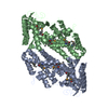
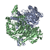
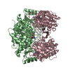
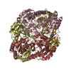
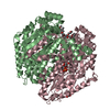
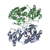
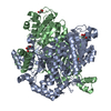
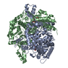
- PDB-2nw9: Crystal Structure of Tryptophan 2,3-dioxygenase (TDO) from Xantho... -
+ Open data

- Basic information
| Entry | Database: PDB / ID: 2nw9 | ||||||
|---|---|---|---|---|---|---|---|
| Title | Crystal Structure of Tryptophan 2,3-dioxygenase (TDO) from Xanthomonas campestris in complex with ferrous heme and 6-fluoro-tryptophan. Northeast Structural Genomics Target XcR13 | ||||||
 Components Components | Tryptophan 2,3-dioxygenase | ||||||
 Keywords Keywords | OXIDOREDUCTASE / all alpha-helical protein / Structural Genomics / PSI-2 / Protein Structure Initiative / Northeast Structural Genomics Consortium / NESG | ||||||
| Function / homology |  Function and homology information Function and homology informationtryptophan 2,3-dioxygenase / : / L-tryptophan 2,3-dioxygenase activity / : / heme binding / metal ion binding Similarity search - Function | ||||||
| Biological species |  Xanthomonas campestris pv. campestris (bacteria) Xanthomonas campestris pv. campestris (bacteria) | ||||||
| Method |  X-RAY DIFFRACTION / SYNCHROTRON / MOLECULAR REPLACEMENT / Resolution: 1.8 Å X-RAY DIFFRACTION / SYNCHROTRON / MOLECULAR REPLACEMENT / Resolution: 1.8 Å | ||||||
 Authors Authors | Forouhar, F. / Anderson, J.L.R. / Mowat, C.G. / Bruckmann, C. / Thackray, S.J. / Seetharaman, J. / Ho, C.K. / Ma, L.C. / Cunningham, K. / Janjua, H. ...Forouhar, F. / Anderson, J.L.R. / Mowat, C.G. / Bruckmann, C. / Thackray, S.J. / Seetharaman, J. / Ho, C.K. / Ma, L.C. / Cunningham, K. / Janjua, H. / Zhao, L. / Xiao, R. / Liu, J. / Baran, M.C. / Acton, T.B. / Rost, B. / Montelione, G.T. / Champman, S.K. / Tong, L. / Northeast Structural Genomics Consortium (NESG) | ||||||
 Citation Citation | Journal: Proc.Natl.Acad.Sci.Usa / Year: 2007 Title: Molecular insights into substrate recognition and catalysis by tryptophan 2,3-dioxygenase. Authors: Forouhar, F. / Anderson, J.L. / Mowat, C.G. / Vorobiev, S.M. / Hussain, A. / Abashidze, M. / Bruckmann, C. / Thackray, S.J. / Seetharaman, J. / Tucker, T. / Xiao, R. / Ma, L.C. / Zhao, L. / ...Authors: Forouhar, F. / Anderson, J.L. / Mowat, C.G. / Vorobiev, S.M. / Hussain, A. / Abashidze, M. / Bruckmann, C. / Thackray, S.J. / Seetharaman, J. / Tucker, T. / Xiao, R. / Ma, L.C. / Zhao, L. / Acton, T.B. / Montelione, G.T. / Chapman, S.K. / Tong, L. | ||||||
| History |
|
- Structure visualization
| Structure viewer | Molecule: MolmilJmol/JSmol |
|---|
- Downloads & links
-Download
| PDBx/mmCIF format | 2nw9.cif.gz | 140.6 KB | Display | PDBx/mmCIF format |
|---|---|---|---|---|
| PDB format | pdb2nw9.ent.gz | 108.7 KB | Display | PDB format |
| PDBx/mmJSON format | 2nw9.json.gz | Tree view | PDBx/mmJSON format | |
| Others |  Other downloads Other downloads |
-Validation report
| Arichive directory | https://data.pdbj.org/pub/pdb/validation_reports/nw/2nw9ftp://data.pdbj.org/pub/pdb/validation_reports/nw/2nw9 | HTTPS FTP |
|---|
-Related structure data
| Related structure data |  2nw7C 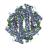 2nw8C  2nwbC  1yw0S S: Starting model for refinement C: citing same article ( |
|---|---|
| Similar structure data | |
| Other databases |


-Links
 PDBj
PDBj- Assembly
| Deposited unit | 
| ||||||||
|---|---|---|---|---|---|---|---|---|---|
| 1 | 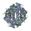
| ||||||||
| Unit cell |
|
-Components
| #1: Protein | Mass: 35731.422 Da / Num. of mol.: 2 Source method: isolated from a genetically manipulated source Source: (gene. exp.) Xanthomonas campestris pv. campestris (bacteria)Species: Xanthomonas campestris / Strain: pv. campestris / Gene: XCC0432 / Plasmid: BL21 / Production host: #2: Chemical | ChemComp-FT6 / | 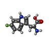  Type: L-peptide linking / Mass: 222.216 Da / Num. of mol.: 1 / Source method: obtained synthetically / Formula: C11H11FN2O2 Type: L-peptide linking / Mass: 222.216 Da / Num. of mol.: 1 / Source method: obtained synthetically / Formula: C11H11FN2O2#3: Chemical |   Mass: 616.487 Da / Num. of mol.: 2 / Source method: obtained synthetically / Formula: C34H32FeN4O4 Mass: 616.487 Da / Num. of mol.: 2 / Source method: obtained synthetically / Formula: C34H32FeN4O4#4: Chemical |   Mass: 54.938 Da / Num. of mol.: 2 / Source method: obtained synthetically / Formula: Mn Mass: 54.938 Da / Num. of mol.: 2 / Source method: obtained synthetically / Formula: Mn#5: Water | ChemComp-HOH / |  Mass: 18.015 Da / Num. of mol.: 596 / Source method: isolated from a natural source / Formula: H2O Mass: 18.015 Da / Num. of mol.: 596 / Source method: isolated from a natural source / Formula: H2O |
|---|
-Experimental details
-Experiment
| Experiment | Method: X-RAY DIFFRACTION |
|---|
- Sample preparation
| Crystal | Density Matthews: 2.53 Å3/Da / Density % sol: 51.4 % |
|---|---|
| Crystal grow | Temperature: 293 K / Method: vapor diffusion, sitting drop / pH: 6.3 Details: 100mM MES, 10% PEG4000, 60mM Manganese chloride, 10mM Sodium dithionite, 8mM 6-fluoro-D/L-tryptophan, pH 6.3, VAPOR DIFFUSION, SITTING DROP, temperature 293K |
-Data collection
| Diffraction | Mean temperature: 100 K |
|---|---|
| Diffraction source | Source: SYNCHROTRON / Site: ESRF  / Beamline: BM14 / Wavelength: 0.979 Å / Beamline: BM14 / Wavelength: 0.979 Å |
| Detector | Type: MARMOSAIC 225 mm CCD / Detector: CCD / Date: Jun 20, 2006 / Details: mirrors |
| Radiation | Monochromator: Si 111 CHANNEL / Protocol: SINGLE WAVELENGTH / Monochromatic (M) / Laue (L): M / Scattering type: x-ray |
| Radiation wavelength | Wavelength: 0.979 Å / Relative weight: 1 |
| Reflection | Resolution: 1.8→26.38 Å / Num. all: 67279 / Num. obs: 67279 / % possible obs: 99.9 % / Observed criterion σ(F): 0 / Observed criterion σ(I): 0 / Redundancy: 7.4 % / Biso Wilson estimate: 15 Å2 / Rmerge(I) obs: 0.086 / Rsym value: 0.066 / Net I/σ(I): 19.97 |
| Reflection shell | Resolution: 1.8→1.86 Å / Redundancy: 7.3 % / Rmerge(I) obs: 0.609 / Mean I/σ(I) obs: 2.5 / Rsym value: 0.56 / % possible all: 100 |
- Processing
| Software |
| ||||||||||||||||||||||||||||||||||||||||||||||||||||||||||||
|---|---|---|---|---|---|---|---|---|---|---|---|---|---|---|---|---|---|---|---|---|---|---|---|---|---|---|---|---|---|---|---|---|---|---|---|---|---|---|---|---|---|---|---|---|---|---|---|---|---|---|---|---|---|---|---|---|---|---|---|---|---|
| Refinement | Method to determine structure: MOLECULAR REPLACEMENT Starting model: PDB entry 1YW0 Resolution: 1.8→26.38 Å / Rfactor Rfree error: 0.003 / Data cutoff high absF: 61158.59 / Data cutoff low absF: 0 / Isotropic thermal model: OVERALL / Cross valid method: THROUGHOUT / σ(F): 2 / Stereochemistry target values: Engh & Huber / Details: XtalView was also the program used in refinement
| ||||||||||||||||||||||||||||||||||||||||||||||||||||||||||||
| Solvent computation | Solvent model: FLAT MODEL / Bsol: 51.7096 Å2 / ksol: 0.35712 e/Å3 | ||||||||||||||||||||||||||||||||||||||||||||||||||||||||||||
| Displacement parameters | Biso mean: 23.8 Å2
| ||||||||||||||||||||||||||||||||||||||||||||||||||||||||||||
| Refine analyze |
| ||||||||||||||||||||||||||||||||||||||||||||||||||||||||||||
| Refinement step | Cycle: LAST / Resolution: 1.8→26.38 Å
| ||||||||||||||||||||||||||||||||||||||||||||||||||||||||||||
| Refine LS restraints |
| ||||||||||||||||||||||||||||||||||||||||||||||||||||||||||||
| LS refinement shell | Resolution: 1.8→1.86 Å / Rfactor Rfree error: 0.015 / Total num. of bins used: 10
|
