 Movie
Movie Controller

[English] 日本語
 Yorodumi
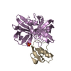
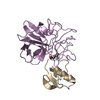
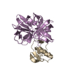
- PDB-2nu3: Accommodation of positively-charged residues in a hydrophobic spe... -
+ Open data

- Basic information
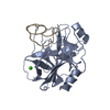
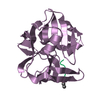
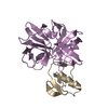
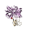
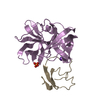
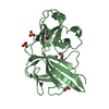
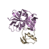
| Entry | Database: PDB / ID: 2nu3 | ||||||
|---|---|---|---|---|---|---|---|
| Title | Accommodation of positively-charged residues in a hydrophobic specificity pocket: Crystal structures of SGPB in complex with OMTKY3 variants Lys18I and Arg18I | ||||||
 Components Components |
| ||||||
 Keywords Keywords | HYDROLASE / ENZYME-INHIBITOR COMPLEX / CHARGED P1 RESIDUE | ||||||
| Function / homology |  Function and homology information Function and homology informationstreptogrisin B / molecular function inhibitor activity / serine-type endopeptidase inhibitor activity / serine-type endopeptidase activity / proteolysis / extracellular region Similarity search - Function | ||||||
| Biological species |   Streptomyces griseus (bacteria) Streptomyces griseus (bacteria) | ||||||
| Method |  X-RAY DIFFRACTION / MIR / Resolution: 1.8 Å X-RAY DIFFRACTION / MIR / Resolution: 1.8 Å | ||||||
 Authors Authors | Bateman, K.S. / Anderson, S. / Lu, W. / Qasim, M.A. / Huang, K. / Laskowski Jr., M. / James, M.N.G. | ||||||
 Citation Citation | Journal: To be Published Title: Accommodation of positively-charged residues in a hydrophobic specificity pocket: Crystal structures of SGPB in complex with OMTKY3 variants Lys18I and Arg18I Authors: Bateman, K.S. / Anderson, S. / Lu, W. / Qasim, M.A. / Huang, K. / Laskowski Jr., M. / James, M.N.G. | ||||||
| History |
|
- Structure visualization
| Structure viewer | Molecule: MolmilJmol/JSmol |
|---|
- Downloads & links
-Download
| PDBx/mmCIF format | 2nu3.cif.gz | 59.2 KB | Display | PDBx/mmCIF format |
|---|---|---|---|---|
| PDB format | pdb2nu3.ent.gz | 41.2 KB | Display | PDB format |
| PDBx/mmJSON format | 2nu3.json.gz | Tree view | PDBx/mmJSON format | |
| Others |  Other downloads Other downloads |
-Validation report
| Arichive directory | https://data.pdbj.org/pub/pdb/validation_reports/nu/2nu3ftp://data.pdbj.org/pub/pdb/validation_reports/nu/2nu3 | HTTPS FTP |
|---|
-Related structure data
| Related structure data |  2nu2C  2nu4C 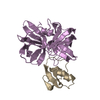 3sgbS S: Starting model for refinement C: citing same article ( |
|---|---|
| Similar structure data |


-Links
 PDBj
PDBj
- Assembly
| Deposited unit | 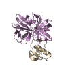
| ||||||||
|---|---|---|---|---|---|---|---|---|---|
| 1 |
| ||||||||
| Unit cell |
|
-Components
| #1: Protein | Mass: 18653.232 Da / Num. of mol.: 1 / Fragment: residues 115-299 / Source method: isolated from a natural source / Source: (natural) Streptomyces griseus (bacteria) / Strain: K1 / References: UniProt: P00777, streptogrisin B |
|---|---|
| #2: Protein | Mass: 5601.311 Da / Num. of mol.: 1 / Fragment: third domain, residues 135-185 / Mutation: L18K Source method: isolated from a genetically manipulated source Source: (gene. exp.) |
| #3: Water | ChemComp-HOH /  Mass: 18.015 Da / Num. of mol.: 151 / Source method: isolated from a natural source / Formula: H2O Mass: 18.015 Da / Num. of mol.: 151 / Source method: isolated from a natural source / Formula: H2O |
| Has protein modification | Y |
-Experimental details
-Experiment
| Experiment | Method: X-RAY DIFFRACTION / Number of used crystals: 1 |
|---|
- Sample preparation
| Crystal | Density Matthews: 2.03 Å3/Da / Density % sol: 39.39 % |
|---|---|
| Crystal grow | Temperature: 298 K / Method: vapor diffusion, hanging drop / pH: 7.1 Details: PEG 4000, SODIUM POTASSIUM PHOSPHATE, pH 7.1, VAPOR DIFFUSION, HANGING DROP, temperature 298K |
-Data collection
| Diffraction | Mean temperature: 298 K |
|---|---|
| Diffraction source | Source: ROTATING ANODE / Type: RIGAKU / Wavelength: 1.54178 Å |
| Detector | Type: SDMS / Detector: AREA DETECTOR / Date: Sep 5, 1995 / Details: graphite monochrometer |
| Radiation | Monochromator: graphite / Protocol: SINGLE WAVELENGTH / Monochromatic (M) / Laue (L): M / Scattering type: x-ray |
| Radiation wavelength | Wavelength: 1.54178 Å / Relative weight: 1 |
| Reflection | Resolution: 1.8→20 Å / Num. obs: 17246 / % possible obs: 94.8 % / Observed criterion σ(F): 0 / Observed criterion σ(I): 2 / Redundancy: 2.7 % / Rmerge(I) obs: 0.056 / Net I/σ(I): 18.27 |
| Reflection shell | Resolution: 1.8→1.94 Å / Rmerge(I) obs: 0.187 / Mean I/σ(I) obs: 2.26 / % possible all: 40.8 |
- Processing
| Software |
| |||||||||||||||||||||
|---|---|---|---|---|---|---|---|---|---|---|---|---|---|---|---|---|---|---|---|---|---|---|
| Refinement | Method to determine structure: MIR Starting model: PDB ENTRY 3SGB Resolution: 1.8→20 Å / σ(F): 0 / σ(I): 0 / Stereochemistry target values: Engh & Huber /
| |||||||||||||||||||||
| Refine analyze | Luzzati coordinate error obs: 0.16 Å / Luzzati sigma a obs: 0.12 Å | |||||||||||||||||||||
| Refinement step | Cycle: LAST / Resolution: 1.8→20 Å
| |||||||||||||||||||||
| Refine LS restraints |
|
