 Movie
Movie Controller

[English] 日本語
 Yorodumi
- PDB-3sgb: STRUCTURE OF THE COMPLEX OF STREPTOMYCES GRISEUS PROTEASE B AND T... -
+ Open data

- Basic information
| Entry | Database: PDB / ID: 3sgb | |||||||||
|---|---|---|---|---|---|---|---|---|---|---|
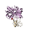
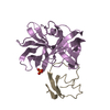
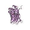
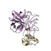
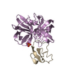
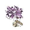
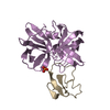
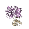
| Title | STRUCTURE OF THE COMPLEX OF STREPTOMYCES GRISEUS PROTEASE B AND THE THIRD DOMAIN OF THE TURKEY OVOMUCOID INHIBITOR AT 1.8 ANGSTROMS RESOLUTION | |||||||||
 Components Components |
| |||||||||
 Keywords Keywords | COMPLEX(SERINE PROTEINASE-INHIBITOR) | |||||||||
| Function / homology |  Function and homology information Function and homology informationstreptogrisin B / molecular function inhibitor activity / serine-type endopeptidase inhibitor activity / serine-type endopeptidase activity / proteolysis / extracellular region Similarity search - Function | |||||||||
| Biological species |  Streptomyces griseus (bacteria) Streptomyces griseus (bacteria) | |||||||||
| Method |  X-RAY DIFFRACTION / Resolution: 1.8 Å X-RAY DIFFRACTION / Resolution: 1.8 Å | |||||||||
 Authors Authors | Read, R.J. / Fujinaga, M. / Sielecki, A.R. / James, M.N.G. | |||||||||
 Citation Citation | Journal: Biochemistry / Year: 1983 Title: Structure of the complex of Streptomyces griseus protease B and the third domain of the turkey ovomucoid inhibitor at 1.8-A resolution. Authors: Read, R.J. / Fujinaga, M. / Sielecki, A.R. / James, M.N. #1: Journal: J.Mol.Biol. / Year: 1987Title: Crystal and Molecular Structures of the Complex of Alpha-Chymotrypsin with its Inhibitor Turkey Ovomucoid Third Domain at 1.8 Angstroms Resolution Authors: Fujinaga, M. / Sielecki, A.R. / Read, R.J. / Ardelt, W. / Laskowskijunior, M. / James, M.N.G. #2: Journal: Proc.Natl.Acad.Sci.USA / Year: 1982Title: Refined Crystal Structure of the Molecular Complex of Streptomyces Griseus Protease B, a Serine Protease, with the Third Domain of the Ovomucoid Inhibitor from Turkey Authors: Fujinaga, M. / Read, R.J. / Sielecki, A. / Ardelt, W. / Laskowski Junior, M. / James, M.N.G. #3: Journal: J.Mol.Biol. / Year: 1980Title: Crystal Structure Studies and Inhibition Kinetics of Tripeptide Chloromethyl Ketone Inhibitors with Streptomyces Griseus Protease B Authors: James, M.N.G. / Brayer, G.D. / Delbaere, L.T.J. / Sielecki, A.R. / Gertler, A. #4: Journal: Can.J.Biochem. / Year: 1979Title: The 2.8 Angstroms Resolution Structure of Streptomyces Griseus Protease B and its Homology with Alpha-Chymotrypsin and Streptomyces Griseus Protease A Authors: Delbaere, L.T.J. / Brayer, G.D. / James, M.M.G. #5: Journal: Can.J.Biochem. / Year: 1978Title: Amino Acid Sequence Alignment of Bacterial and Mammalian Pancreatic Serine Proteases Based on Topological Equivalences Authors: James, M.N.G. / Delbaere, L.T.J. / Brayer, G.D. #6: Journal: Miami Winter Symp. / Year: 1976Title: Relationship between the Structures and Activities of Some Microbial Serine Proteases. II. Comparison of the Tertiary Structures of Microbial and Pancreatic Serine Proteases Authors: James, M.N.G. #7: Journal: Nature / Year: 1975Title: Tertiary Structural Differences between Microbial Serine Proteases and Pancreatic Serine Enzymes Authors: Delbaere, L.T.J. / Hutcheon, W.L.B. / James, M.N.G. / Theissen, W.E. #8: Journal: Can.J.Biochem. / Year: 1974Title: 4.5 Angstroms Resolution Structure of a Bacterial Serine Protease from Streptomyces Griseus Authors: Codding, P.W. / Delbaere, L.T.J. / Hayakawa, K. / Hutcheon, W.L.B. / James, M.N.G. / Jurasek, L. | |||||||||
| History |
| |||||||||
| Remark 700 | SHEET THE SEVEN-STRANDED SHEETS BL1 AND BL2 PRESENTED ON SHEET RECORDS BELOW ARE ACTUALLY SIX- ...SHEET THE SEVEN-STRANDED SHEETS BL1 AND BL2 PRESENTED ON SHEET RECORDS BELOW ARE ACTUALLY SIX-STRANDED BETA BARRELS. THIS IS DENOTED BY THE FIRST STRAND RECURRING AS THE LAST STRAND. |
- Structure visualization
| Structure viewer | Molecule: MolmilJmol/JSmol |
|---|
- Downloads & links
-Download
| PDBx/mmCIF format | 3sgb.cif.gz | 59.7 KB | Display | PDBx/mmCIF format |
|---|---|---|---|---|
| PDB format | pdb3sgb.ent.gz | 42.8 KB | Display | PDB format |
| PDBx/mmJSON format | 3sgb.json.gz | Tree view | PDBx/mmJSON format | |
| Others |  Other downloads Other downloads |
-Validation report
| Arichive directory | https://data.pdbj.org/pub/pdb/validation_reports/sg/3sgbftp://data.pdbj.org/pub/pdb/validation_reports/sg/3sgb | HTTPS FTP |
|---|
-Related structure data
| Similar structure data |
|---|


-Links
 PDBj
PDBj
- Assembly
| Deposited unit | 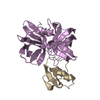
| ||||||||
|---|---|---|---|---|---|---|---|---|---|
| 1 |
| ||||||||
| Unit cell |
|
-Components
| #1: Protein | Mass: 18665.285 Da / Num. of mol.: 1 Source method: isolated from a genetically manipulated source Source: (gene. exp.) Streptomyces griseus (bacteria) / Organ: PANCREATIC / References: UniProt: P00777 |
|---|---|
| #2: Protein | Mass: 6026.811 Da / Num. of mol.: 1 Source method: isolated from a genetically manipulated source References: UniProt: P68390 |
| #3: Water | ChemComp-HOH /  Mass: 18.015 Da / Num. of mol.: 182 / Source method: isolated from a natural source / Formula: H2O Mass: 18.015 Da / Num. of mol.: 182 / Source method: isolated from a natural source / Formula: H2O |
| Has protein modification | Y |
-Experimental details
-Experiment
| Experiment | Method: X-RAY DIFFRACTION |
|---|
- Sample preparation
| Crystal | Density Matthews: 1.99 Å3/Da / Density % sol: 38.33 % | |||||||||||||||||||||||||||||||||||
|---|---|---|---|---|---|---|---|---|---|---|---|---|---|---|---|---|---|---|---|---|---|---|---|---|---|---|---|---|---|---|---|---|---|---|---|---|
| Crystal grow | *PLUS pH: 6 / Method: vapor diffusion | |||||||||||||||||||||||||||||||||||
| Components of the solutions | *PLUS
|
-Data collection
| Reflection | *PLUS Highest resolution: 1.8 Å / Num. all: 20227 / Num. obs: 18082 / Num. measured all: 13937 / Rmerge(I) obs: 2 / Biso Wilson estimate: 13.1 Å2 |
|---|
- Processing
| Refinement | Resolution: 1.8→6 Å / Rfactor Rwork: 0.125 | ||||||||||||||||||||||||
|---|---|---|---|---|---|---|---|---|---|---|---|---|---|---|---|---|---|---|---|---|---|---|---|---|---|
| Refine analyze | Luzzati coordinate error obs: 0.14 Å | ||||||||||||||||||||||||
| Refinement step | Cycle: LAST / Resolution: 1.8→6 Å
| ||||||||||||||||||||||||
| Refinement | *PLUS Num. reflection obs: 13457 / σ(I): 2 / Highest resolution: 1.8 Å / Lowest resolution: 6 Å / Rfactor obs: 0.125 | ||||||||||||||||||||||||
| Solvent computation | *PLUS | ||||||||||||||||||||||||
| Displacement parameters | *PLUS | ||||||||||||||||||||||||
| Refine LS restraints | *PLUS
|
