 Movie
Movie Controller

[English] 日本語
 Yorodumi
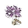
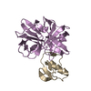
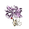
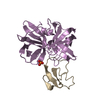
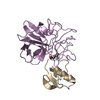
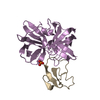
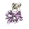
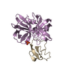
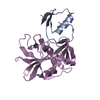
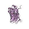
- PDB-1sgn: ASN 18 VARIANT OF TURKEY OVOMUCOID INHIBITOR THIRD DOMAIN COMPLEX... -
+ Open data

- Basic information
| Entry | Database: PDB / ID: 1sgn | ||||||
|---|---|---|---|---|---|---|---|
| Title | ASN 18 VARIANT OF TURKEY OVOMUCOID INHIBITOR THIRD DOMAIN COMPLEXED WITH STREPTOMYCES GRISEUS PROTEINASE B | ||||||
 Components Components |
| ||||||
 Keywords Keywords | HYDROLASE/HYDROLASE INHIBITOR / COMPLEX (SERINE PROTEASE-INHIBITOR) / SERINE PROTEINASE / PROTEIN INHIBITOR / HYDROLASE-HYDROLASE INHIBITOR COMPLEX | ||||||
| Function / homology |  Function and homology information Function and homology informationstreptogrisin B / molecular function inhibitor activity / serine-type endopeptidase inhibitor activity / serine-type endopeptidase activity / proteolysis / extracellular region Similarity search - Function | ||||||
| Biological species |   Streptomyces griseus (bacteria) Streptomyces griseus (bacteria) | ||||||
| Method |  X-RAY DIFFRACTION / MOLECULAR REPLACEMENT / Resolution: 1.8 Å X-RAY DIFFRACTION / MOLECULAR REPLACEMENT / Resolution: 1.8 Å | ||||||
 Authors Authors | Huang, K. / Lu, W. / Anderson, S. / Laskowski Jr., M. / James, M.N.G. | ||||||
 Citation Citation | Journal: To be Published Title: Recruitment of a Buried K+ Ion to Stabilize the Negative Charge of Ionized P1 in the Hydrophobic Pocket: Crystal Structures of Glu18, Gln18, Asp18 and Asn18 Variants of Turkey Ovomucoid ...Title: Recruitment of a Buried K+ Ion to Stabilize the Negative Charge of Ionized P1 in the Hydrophobic Pocket: Crystal Structures of Glu18, Gln18, Asp18 and Asn18 Variants of Turkey Ovomucoid Inhibitor Third Domain Complexed with Streptomyces griseus Protease B at Various pHs Authors: Huang, K. / Lu, W. / Anderson, S. / Laskowski Jr., M. / James, M.N.G. #1: Journal: Protein Sci. / Year: 1995Title: Water molecules participate in proteinase-inhibitor interactions: crystal structures of Leu18, Ala18, and Gly18 variants of turkey ovomucoid inhibitor third domain complexed with Streptomyces griseus proteinase B. Authors: Huang, K. / Lu, W. / Anderson, S. / Laskowski, M. / James, M.N. #2: Journal: Biochemistry / Year: 1983Title: Structure of the Complex of Streptomyces griseus Protease B and the Third Domain of the Turkey Ovomucoid Inhibitor at 1.8 Angstroms Resolution Authors: Read, R.J. / Fujinaga, M. / Sielecki, A.R. / James, M.N.G. | ||||||
| History |
|
- Structure visualization
| Structure viewer | Molecule: MolmilJmol/JSmol |
|---|
- Downloads & links
-Download
| PDBx/mmCIF format | 1sgn.cif.gz | 60.3 KB | Display | PDBx/mmCIF format |
|---|---|---|---|---|
| PDB format | pdb1sgn.ent.gz | 42.3 KB | Display | PDB format |
| PDBx/mmJSON format | 1sgn.json.gz | Tree view | PDBx/mmJSON format | |
| Others |  Other downloads Other downloads |
-Validation report
| Arichive directory | https://data.pdbj.org/pub/pdb/validation_reports/sg/1sgnftp://data.pdbj.org/pub/pdb/validation_reports/sg/1sgn | HTTPS FTP |
|---|
-Related structure data
| Related structure data |  1sgeC  2sgdC  2sgfC 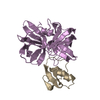 3sgbS S: Starting model for refinement C: citing same article ( |
|---|---|
| Similar structure data |


-Links
 PDBj
PDBj
- Assembly
| Deposited unit | 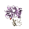
| ||||||||
|---|---|---|---|---|---|---|---|---|---|
| 1 |
| ||||||||
| Unit cell |
|
-Components
| #1: Protein | Mass: 18653.232 Da / Num. of mol.: 1 / Source method: isolated from a natural source / Source: (natural) Streptomyces griseus (bacteria) / Strain: K1 / References: UniProt: P00777, streptogrisin B |
|---|---|
| #2: Protein | Mass: 5586.233 Da / Num. of mol.: 1 / Fragment: THIRD DOMAIN / Mutation: DEL(1-5), L18N Source method: isolated from a genetically manipulated source Source: (gene. exp.) |
| #3: Chemical | ChemComp-PO4 /   Mass: 94.971 Da / Num. of mol.: 1 / Source method: obtained synthetically / Formula: PO4 Mass: 94.971 Da / Num. of mol.: 1 / Source method: obtained synthetically / Formula: PO4 |
| #4: Water | ChemComp-HOH /  Mass: 18.015 Da / Num. of mol.: 161 / Source method: isolated from a natural source / Formula: H2O Mass: 18.015 Da / Num. of mol.: 161 / Source method: isolated from a natural source / Formula: H2O |
| Has protein modification | Y |
-Experimental details
-Experiment
| Experiment | Method: X-RAY DIFFRACTION / Number of used crystals: 1 |
|---|
- Sample preparation
| Crystal | Density Matthews: 2.05 Å3/Da / Density % sol: 39.89 % |
|---|---|
| Crystal grow | pH: 6.5 Details: 2% PEG 6000, 50MM SODIUM/POTASSIUM PHOSPHATE BUFFER AT PH 6.5, pH 6.50 |
-Data collection
| Diffraction | Mean temperature: 287 K |
|---|---|
| Diffraction source | Source: ROTATING ANODE / Type: RIGAKU RU200 / Wavelength: 1.5418 |
| Detector | Type: SDMS / Detector: AREA DETECTOR |
| Radiation | Monochromator: NI FILTER / Protocol: SINGLE WAVELENGTH / Monochromatic (M) / Laue (L): M / Scattering type: x-ray |
| Radiation wavelength | Wavelength: 1.5418 Å / Relative weight: 1 |
| Reflection | Resolution: 1.8→20 Å / Num. obs: 17283 / % possible obs: 89.2 % / Redundancy: 2.7 % / Rmerge(I) obs: 0.07 |
- Processing
| Software | Name: TNT / Classification: refinement | ||||||||||||||||||||||||||||||
|---|---|---|---|---|---|---|---|---|---|---|---|---|---|---|---|---|---|---|---|---|---|---|---|---|---|---|---|---|---|---|---|
| Refinement | Method to determine structure: MOLECULAR REPLACEMENT Starting model: 3SGB Resolution: 1.8→20 Å / σ(F): 0 / Stereochemistry target values: TNT PROTGEO
| ||||||||||||||||||||||||||||||
| Refinement step | Cycle: LAST / Resolution: 1.8→20 Å
| ||||||||||||||||||||||||||||||
| Refine LS restraints |
|
