 Movie
Movie Controller


+ Open data

- Basic information
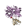
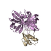
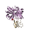
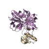
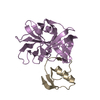
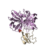
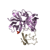
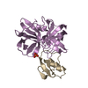
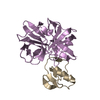
| Entry | Database: PDB / ID: 1ds2 | ||||||
|---|---|---|---|---|---|---|---|
| Title | CRYSTAL STRUCTURE OF SGPB:OMTKY3-COO-LEU18I | ||||||
 Components Components |
| ||||||
 Keywords Keywords | HYDROLASE/HYDROLASE INHIBITOR / serine proteinase / protein inhibitor / ovomucoid / canonical inhibitor / ester bond / SGPB / OMTKY3 / HYDROLASE-HYDROLASE INHIBITOR COMPLEX | ||||||
| Function / homology |  Function and homology information Function and homology informationstreptogrisin B / molecular function inhibitor activity / serine-type endopeptidase inhibitor activity / serine-type endopeptidase activity / proteolysis / extracellular region Similarity search - Function | ||||||
| Biological species |   Streptomyces griseus (bacteria) Streptomyces griseus (bacteria) | ||||||
| Method |  X-RAY DIFFRACTION / Resolution: 1.7 Å X-RAY DIFFRACTION / Resolution: 1.7 Å | ||||||
 Authors Authors | Bateman, K.S. / Huang, K. / Anderson, S. / Lu, W. / Qasim, M.A. / Laskowski Jr., M. / James, M.N.G. | ||||||
 Citation Citation | Journal: J.Mol.Biol. / Year: 2001 Title: Contribution of peptide bonds to inhibitor-protease binding: crystal structures of the turkey ovomucoid third domain backbone variants OMTKY3-Pro18I and OMTKY3-psi[COO]-Leu18I in complex with ...Title: Contribution of peptide bonds to inhibitor-protease binding: crystal structures of the turkey ovomucoid third domain backbone variants OMTKY3-Pro18I and OMTKY3-psi[COO]-Leu18I in complex with Streptomyces griseus proteinase B (SGPB) and the structure of the free inhibitor, OMTKY-3-psi[CH2NH2+]-Asp19I Authors: Bateman, K.S. / Huang, K. / Anderson, S. / Lu, W. / Qasim, M.A. / Laskowski Jr., M. / James, M.N. | ||||||
| History |
|
- Structure visualization
| Structure viewer | Molecule: MolmilJmol/JSmol |
|---|
- Downloads & links
-Download
| PDBx/mmCIF format | 1ds2.cif.gz | 54.9 KB | Display | PDBx/mmCIF format |
|---|---|---|---|---|
| PDB format | pdb1ds2.ent.gz | 41.6 KB | Display | PDB format |
| PDBx/mmJSON format | 1ds2.json.gz | Tree view | PDBx/mmJSON format | |
| Others |  Other downloads Other downloads |
-Validation report
| Arichive directory | https://data.pdbj.org/pub/pdb/validation_reports/ds/1ds2ftp://data.pdbj.org/pub/pdb/validation_reports/ds/1ds2 | HTTPS FTP |
|---|
-Related structure data


-Links
 PDBj
PDBj
- Assembly
| Deposited unit | 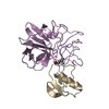
| ||||||||
|---|---|---|---|---|---|---|---|---|---|
| 1 |
| ||||||||
| Unit cell |
|
-Components
| #1: Protein | Mass: 18665.285 Da / Num. of mol.: 1 / Source method: isolated from a natural source / Source: (natural) Streptomyces griseus (bacteria) / References: UniProt: P00777, streptogrisin B |
|---|---|
| #2: Protein | Mass: 5586.273 Da / Num. of mol.: 1 / Fragment: THIRD DOMAIN (OMTKY3) / Mutation: ESTER BOND BETWEEN THR17I-LEU18I(1LU) Source method: isolated from a genetically manipulated source Source: (gene. exp.) Description: THE PEPTIDE LINKAGE BETWEEN THR17I AND LEU18I(1LU) HAS BEEN REPLACED BY AN ESTER BOND. Production host: |
| #3: Water | ChemComp-HOH /  Mass: 18.015 Da / Num. of mol.: 140 / Source method: isolated from a natural source / Formula: H2O Mass: 18.015 Da / Num. of mol.: 140 / Source method: isolated from a natural source / Formula: H2O |
-Experimental details
-Experiment
| Experiment | Method: X-RAY DIFFRACTION / Number of used crystals: 1 |
|---|
- Sample preparation
| Crystal | Density Matthews: 2.03 Å3/Da / Density % sol: 39.53 % | ||||||||||||||||||||||||||||
|---|---|---|---|---|---|---|---|---|---|---|---|---|---|---|---|---|---|---|---|---|---|---|---|---|---|---|---|---|---|
| Crystal grow | Temperature: 298 K / Method: vapor diffusion, hanging drop / pH: 7.1 Details: PEG 4000, sodium potassium phosphate, pH 7.1, VAPOR DIFFUSION, HANGING DROP, temperature 298.0K | ||||||||||||||||||||||||||||
| Crystal grow | *PLUS Details: used seeding / PH range low: 7.1 / PH range high: 6.5 | ||||||||||||||||||||||||||||
| Components of the solutions | *PLUS
|
-Data collection
| Diffraction | Mean temperature: 298 K |
|---|---|
| Diffraction source | Source: ROTATING ANODE / Type: RIGAKU / Wavelength: 1.5417 |
| Detector | Type: MACSCIENCE / Detector: IMAGE PLATE / Date: Mar 12, 1999 |
| Radiation | Protocol: SINGLE WAVELENGTH / Monochromatic (M) / Laue (L): M / Scattering type: x-ray |
| Radiation wavelength | Wavelength: 1.5417 Å / Relative weight: 1 |
| Reflection | Resolution: 1.7→20 Å / Num. obs: 61078 / % possible obs: 87.8 % / Redundancy: 3.2 % / Rmerge(I) obs: 0.12 / Net I/σ(I): 7.86 |
| Reflection shell | Resolution: 1.7→1.74 Å / Rmerge(I) obs: 0.288 / % possible all: 47.1 |
| Reflection | *PLUS Num. obs: 18894 / Num. measured all: 61078 / Rmerge(I) obs: 0.12 |
| Reflection shell | *PLUS Highest resolution: 1.71 Å / % possible obs: 47.1 % |
- Processing
| Software |
| |||||||||||||||||||||
|---|---|---|---|---|---|---|---|---|---|---|---|---|---|---|---|---|---|---|---|---|---|---|
| Refinement | Resolution: 1.7→20 Å / Stereochemistry target values: Engh & Huber /
| |||||||||||||||||||||
| Refinement step | Cycle: LAST / Resolution: 1.7→20 Å
| |||||||||||||||||||||
| Refine LS restraints |
| |||||||||||||||||||||
| Software | *PLUS Name: TNT / Version: 5E / Classification: refinement | |||||||||||||||||||||
| Refinement | *PLUS Lowest resolution: 20 Å / Rfactor all: 0.183 | |||||||||||||||||||||
| Solvent computation | *PLUS | |||||||||||||||||||||
| Displacement parameters | *PLUS | |||||||||||||||||||||
| Refine LS restraints | *PLUS Type: t_plane_restr / Dev ideal: 0.012 |
