 Movie
Movie Controller


+ Open data

- Basic information
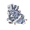
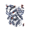
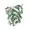
| Entry | Database: PDB / ID: 2nt1 | ||||||
|---|---|---|---|---|---|---|---|
| Title | Structure of acid-beta-glucosidase at neutral pH | ||||||
 Components Components | Glucosylceramidase | ||||||
 Keywords Keywords | HYDROLASE / acid-beta-glucosidase / cerezyme / glucosylceramide / gaucher disease | ||||||
| Function / homology |  Function and homology information Function and homology informationsteryl-beta-glucosidase activity / beta-glucoside catabolic process / cerebellar Purkinje cell layer formation / positive regulation of neuronal action potential / termination of signal transduction / galactosylceramidase / galactosylceramidase activity / glucosylceramidase / lymphocyte migration / regulation of lysosomal protein catabolic process ...steryl-beta-glucosidase activity / beta-glucoside catabolic process / cerebellar Purkinje cell layer formation / positive regulation of neuronal action potential / termination of signal transduction / galactosylceramidase / galactosylceramidase activity / glucosylceramidase / lymphocyte migration / regulation of lysosomal protein catabolic process / scavenger receptor binding / glucosylceramide catabolic process / glycolipid biosynthetic process / sphingosine biosynthetic process / response to thyroid hormone / autophagosome organization / glucosylceramidase activity / lysosomal protein catabolic process / microglial cell proliferation / glucosyltransferase activity / Glycosphingolipid catabolism / lipid storage / microglia differentiation / regulation of TOR signaling / positive regulation of type 2 mitophagy / brain morphogenesis / response to pH / Hydrolases; Glycosylases; Glycosidases, i.e. enzymes that hydrolyse O- and S-glycosyl compounds / pyramidal neuron differentiation / ceramide biosynthetic process / negative regulation of protein metabolic process / Transferases; Glycosyltransferases; Hexosyltransferases / antigen processing and presentation / response to dexamethasone / beta-glucosidase activity / lysosome organization / neuromuscular process / response to testosterone / hematopoietic stem cell proliferation / Association of TriC/CCT with target proteins during biosynthesis / motor behavior / negative regulation of interleukin-6 production / establishment of skin barrier / homeostasis of number of cells / regulation of macroautophagy / cholesterol metabolic process / negative regulation of protein-containing complex assembly / mitophagy / negative regulation of MAPK cascade / cell maturation / lysosomal lumen / thymus development / cellular response to starvation / determination of adult lifespan / respiratory electron transport chain / trans-Golgi network / response to estrogen / negative regulation of inflammatory response / cellular response to tumor necrosis factor / autophagy / T cell differentiation in thymus / positive regulation of proteasomal ubiquitin-dependent protein catabolic process / neuron apoptotic process / negative regulation of neuron apoptotic process / proteasome-mediated ubiquitin-dependent protein catabolic process / lysosome / immune response / signaling receptor binding / lysosomal membrane / Golgi apparatus / endoplasmic reticulum / extracellular exosome Similarity search - Function | ||||||
| Biological species |  Homo sapiens (human) Homo sapiens (human) | ||||||
| Method |  X-RAY DIFFRACTION / SYNCHROTRON / MOLECULAR REPLACEMENT / Resolution: 2.3 Å X-RAY DIFFRACTION / SYNCHROTRON / MOLECULAR REPLACEMENT / Resolution: 2.3 Å | ||||||
 Authors Authors | Lieberman, R.L. / Petsko, G.A. / Ringe, D. | ||||||
 Citation Citation | Journal: Nat.Chem.Biol. / Year: 2007 Title: Structure of acid beta-glucosidase with pharmacological chaperone provides insight into Gaucher disease. Authors: Lieberman, R.L. / Wustman, B.A. / Huertas, P. / Powe, A.C. / Pine, C.W. / Khanna, R. / Schlossmacher, M.G. / Ringe, D. / Petsko, G.A. | ||||||
| History |
|
- Structure visualization
| Structure viewer | Molecule: MolmilJmol/JSmol |
|---|
- Downloads & links
-Download
| PDBx/mmCIF format | 2nt1.cif.gz | 431.9 KB | Display | PDBx/mmCIF format |
|---|---|---|---|---|
| PDB format | pdb2nt1.ent.gz | 353.3 KB | Display | PDB format |
| PDBx/mmJSON format | 2nt1.json.gz | Tree view | PDBx/mmJSON format | |
| Others |  Other downloads Other downloads |
-Validation report
| Arichive directory | https://data.pdbj.org/pub/pdb/validation_reports/nt/2nt1ftp://data.pdbj.org/pub/pdb/validation_reports/nt/2nt1 | HTTPS FTP |
|---|
-Related structure data
| Related structure data |  2nsxC  2nt0SC C: citing same article ( S: Starting model for refinement |
|---|---|
| Similar structure data |


-Links
 PDBj
PDBj


- Assembly
| Deposited unit | 
| ||||||||||||||||||||||||||||||||||||||
|---|---|---|---|---|---|---|---|---|---|---|---|---|---|---|---|---|---|---|---|---|---|---|---|---|---|---|---|---|---|---|---|---|---|---|---|---|---|---|---|
| 1 | 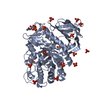
| ||||||||||||||||||||||||||||||||||||||
| 2 | 
| ||||||||||||||||||||||||||||||||||||||
| 3 | 
| ||||||||||||||||||||||||||||||||||||||
| 4 | 
| ||||||||||||||||||||||||||||||||||||||
| 5 |
| ||||||||||||||||||||||||||||||||||||||
| Unit cell |
| ||||||||||||||||||||||||||||||||||||||
| Noncrystallographic symmetry (NCS) | NCS domain:
NCS domain segments: Component-ID: 1 / Beg auth comp-ID: ALA / Beg label comp-ID: ALA / End auth comp-ID: GLN / End label comp-ID: GLN / Refine code: 5 / Auth seq-ID: 1 - 497 / Label seq-ID: 1 - 497
NCS ensembles :
|
-Components
| #1: Protein | Mass: 55640.168 Da / Num. of mol.: 4 Source method: isolated from a genetically manipulated source Source: (gene. exp.) Homo sapiens (human) / Gene: GBA / Organ (production host): Ovary / Production host:   Cricetulus griseus (Chinese hamster) / References: UniProt: P04062, glucosylceramidase Cricetulus griseus (Chinese hamster) / References: UniProt: P04062, glucosylceramidase#2: Sugar | ChemComp-NAG /   Type: D-saccharide, beta linking / Mass: 221.208 Da / Num. of mol.: 4 Type: D-saccharide, beta linking / Mass: 221.208 Da / Num. of mol.: 4Source method: isolated from a genetically manipulated source Formula: C8H15NO6 #3: Chemical | ChemComp-PO4 /   Mass: 94.971 Da / Num. of mol.: 78 / Source method: obtained synthetically / Formula: PO4 Mass: 94.971 Da / Num. of mol.: 78 / Source method: obtained synthetically / Formula: PO4#4: Water | ChemComp-HOH / |  Mass: 18.015 Da / Num. of mol.: 1344 / Source method: isolated from a natural source / Formula: H2O Mass: 18.015 Da / Num. of mol.: 1344 / Source method: isolated from a natural source / Formula: H2OHas protein modification | Y | |
|---|
-Experimental details
-Experiment
| Experiment | Method: X-RAY DIFFRACTION / Number of used crystals: 1 |
|---|
- Sample preparation
| Crystal | Density Matthews: 3.25 Å3/Da / Density % sol: 62.11 % |
|---|---|
| Crystal grow | Temperature: 298 K / Method: vapor diffusion, hanging drop / pH: 7.5 Details: 0.8 M Na dihydrogen phosphate 0.8 M K dihydrogen phosphate 0.1 M Hepes buffer pH 7.5 Cryoprotected in 2 M Lithium sulfate, VAPOR DIFFUSION, HANGING DROP, temperature 298K |
-Data collection
| Diffraction | Mean temperature: 100 K |
|---|---|
| Diffraction source | Source: SYNCHROTRON / Site: APS  / Beamline: 23-ID-B / Wavelength: 1 Å / Beamline: 23-ID-B / Wavelength: 1 Å |
| Detector | Type: MARRESEARCH / Detector: CCD / Date: Jun 30, 2006 |
| Radiation | Protocol: SINGLE WAVELENGTH / Monochromatic (M) / Laue (L): M / Scattering type: x-ray |
| Radiation wavelength | Wavelength: 1 Å / Relative weight: 1 |
| Reflection | Resolution: 2.3→20 Å / Num. obs: 119558 / % possible obs: 95.1 % / Redundancy: 3.5 % / Biso Wilson estimate: 29.1 Å2 / Rsym value: 0.106 / Χ2: 1.432 / Net I/σ(I): 7.8 |
| Reflection shell | Resolution: 2.3→2.38 Å / Redundancy: 2.9 % / Rmerge(I) obs: 0.454 / Mean I/σ(I) obs: 2.4 / Num. unique all: 9993 / Χ2: 1.114 / % possible all: 79.6 |
- Processing
| Software |
| ||||||||||||||||||||||||||||||||||||||||||||||||||||||||||||||||||||||||||||||||||||||||||
|---|---|---|---|---|---|---|---|---|---|---|---|---|---|---|---|---|---|---|---|---|---|---|---|---|---|---|---|---|---|---|---|---|---|---|---|---|---|---|---|---|---|---|---|---|---|---|---|---|---|---|---|---|---|---|---|---|---|---|---|---|---|---|---|---|---|---|---|---|---|---|---|---|---|---|---|---|---|---|---|---|---|---|---|---|---|---|---|---|---|---|---|
| Refinement | Method to determine structure: MOLECULAR REPLACEMENT Starting model: 2NT0 Resolution: 2.3→19.94 Å / Cor.coef. Fo:Fc: 0.953 / Cor.coef. Fo:Fc free: 0.912 / SU B: 6.679 / SU ML: 0.16 / Cross valid method: THROUGHOUT / σ(F): 0 / ESU R: 0.265 / ESU R Free: 0.225 / Stereochemistry target values: MAXIMUM LIKELIHOOD / Details: HYDROGENS HAVE BEEN ADDED IN THE RIDING POSITIONS
| ||||||||||||||||||||||||||||||||||||||||||||||||||||||||||||||||||||||||||||||||||||||||||
| Solvent computation | Ion probe radii: 0.8 Å / Shrinkage radii: 0.8 Å / VDW probe radii: 1.2 Å / Solvent model: MASK | ||||||||||||||||||||||||||||||||||||||||||||||||||||||||||||||||||||||||||||||||||||||||||
| Displacement parameters | Biso mean: 29.096 Å2
| ||||||||||||||||||||||||||||||||||||||||||||||||||||||||||||||||||||||||||||||||||||||||||
| Refinement step | Cycle: LAST / Resolution: 2.3→19.94 Å
| ||||||||||||||||||||||||||||||||||||||||||||||||||||||||||||||||||||||||||||||||||||||||||
| Refine LS restraints |
| ||||||||||||||||||||||||||||||||||||||||||||||||||||||||||||||||||||||||||||||||||||||||||
| Refine LS restraints NCS | Dom-ID: 1 / Refine-ID: X-RAY DIFFRACTION
| ||||||||||||||||||||||||||||||||||||||||||||||||||||||||||||||||||||||||||||||||||||||||||
| LS refinement shell | Resolution: 2.303→2.362 Å / Total num. of bins used: 20
|
