 Movie
Movie Controller

[English] 日本語
 Yorodumi
- PDB-2nr0: Crystal structure of pseudoudirinde synthase TruA in complex with... -
+ Open data

- Basic information
| Entry | Database: PDB / ID: 2nr0 | ||||||
|---|---|---|---|---|---|---|---|
| Title | Crystal structure of pseudoudirinde synthase TruA in complex with leucyl tRNA | ||||||
 Components Components |
| ||||||
 Keywords Keywords | ISOMERASE/RNA / pseudouridine synthase / anticodon stem loop / tRNA / multisite specificity / ISOMERASE-RNA COMPLEX | ||||||
| Function / homology |  Function and homology information Function and homology informationtRNA pseudouridine38-40 synthase / tRNA pseudouridine(38-40) synthase activity / tRNA pseudouridine synthase activity / tRNA pseudouridine synthesis / pseudouridine synthase activity / tRNA binding / protein homodimerization activity Similarity search - Function | ||||||
| Biological species |  | ||||||
| Method |  X-RAY DIFFRACTION / SYNCHROTRON / MOLECULAR REPLACEMENT / Resolution: 3.9 Å X-RAY DIFFRACTION / SYNCHROTRON / MOLECULAR REPLACEMENT / Resolution: 3.9 Å | ||||||
 Authors Authors | Hur, S. / Stroud, R.M. | ||||||
 Citation Citation | Journal: Mol.Cell / Year: 2007 Title: How U38, 39, and 40 of Many tRNAs Become the Targets for Pseudouridylation by TruA. Authors: Hur, S. / Stroud, R.M. | ||||||
| History |
|
- Structure visualization
| Structure viewer | Molecule: MolmilJmol/JSmol |
|---|
- Downloads & links
-Download
| PDBx/mmCIF format | 2nr0.cif.gz | 361 KB | Display | PDBx/mmCIF format |
|---|---|---|---|---|
| PDB format | pdb2nr0.ent.gz | 285.3 KB | Display | PDB format |
| PDBx/mmJSON format | 2nr0.json.gz | Tree view | PDBx/mmJSON format | |
| Others |  Other downloads Other downloads |
-Validation report
| Arichive directory | https://data.pdbj.org/pub/pdb/validation_reports/nr/2nr0ftp://data.pdbj.org/pub/pdb/validation_reports/nr/2nr0 | HTTPS FTP |
|---|
-Related structure data
| Related structure data | 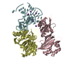 2nqpC  2nreC 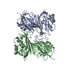 1dj0S 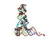 1ehzS S: Starting model for refinement C: citing same article ( |
|---|---|
| Similar structure data |


-Links
 PDBj
PDBj


- Assembly
| Deposited unit | 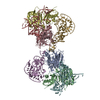
| ||||||||
|---|---|---|---|---|---|---|---|---|---|
| 1 | 
| ||||||||
| 2 | 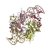
| ||||||||
| Unit cell |
| ||||||||
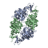
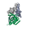
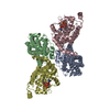
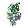
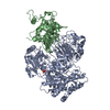
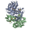
| Details | Molecules of id A & B form a dimeric TruA, and E and F are the substrate tRNAs bound to them.Molecules of id C & D form a dimeric TruA, and G and H are the substrate tRNAs bound to them. |
-Components
| #1: RNA chain | Mass: 28094.645 Da / Num. of mol.: 4 / Source method: obtained synthetically Details: Occurs naturally in E. coli. tRNA is obtained by in vitro transcription. #2: Protein | Mass: 30441.615 Da / Num. of mol.: 4 Source method: isolated from a genetically manipulated source Source: (gene. exp.) References: UniProt: P07649, tRNA pseudouridine38-40 synthase |
|---|
-Experimental details
-Experiment
| Experiment | Method: X-RAY DIFFRACTION / Number of used crystals: 1 |
|---|
- Sample preparation
| Crystal | Density Matthews: 3.17 Å3/Da / Density % sol: 61.18 % | ||||||||||||||||||||||||||||
|---|---|---|---|---|---|---|---|---|---|---|---|---|---|---|---|---|---|---|---|---|---|---|---|---|---|---|---|---|---|
| Crystal grow | Temperature: 298 K / Method: vapor diffusion, hanging drop / pH: 7.5 Details: 20% PEG 3350, 0.2 M K3 Citrate, pH 7.5, VAPOR DIFFUSION, HANGING DROP, temperature 298K | ||||||||||||||||||||||||||||
| Components of the solutions |
|
-Data collection
| Diffraction | Mean temperature: 100 K |
|---|---|
| Diffraction source | Source: SYNCHROTRON / Site: ALS  / Beamline: 8.3.1 / Wavelength: 1 Å / Beamline: 8.3.1 / Wavelength: 1 Å |
| Detector | Type: ADSC QUANTUM 210 / Detector: CCD / Date: Dec 12, 2004 |
| Radiation | Monochromator: Double crystal Si(111) / Protocol: SINGLE WAVELENGTH / Monochromatic (M) / Laue (L): M / Scattering type: x-ray |
| Radiation wavelength | Wavelength: 1 Å / Relative weight: 1 |
| Reflection | Resolution: 3.9→50 Å / Num. all: 24919 / Num. obs: 24919 / % possible obs: 93.3 % / Observed criterion σ(F): 0 / Observed criterion σ(I): 0 / Redundancy: 2.8 % / Rsym value: 0.213 / Net I/σ(I): 4.86 |
| Reflection shell | Resolution: 3.9→4.04 Å / Redundancy: 2.6 % / Mean I/σ(I) obs: 1.56 / Num. unique all: 2453 / Rsym value: 0.619 / % possible all: 93 |
- Processing
| Software |
| |||||||||||||||||||||||||
|---|---|---|---|---|---|---|---|---|---|---|---|---|---|---|---|---|---|---|---|---|---|---|---|---|---|---|
| Refinement | Method to determine structure: MOLECULAR REPLACEMENT Starting model: 1DJ0 (apo TruA enzyme) and 1EHZ (phe tRNA) Resolution: 3.9→50 Å / Isotropic thermal model: MAXIMUM LIKELIHOOD / Cross valid method: THROUGHOUT / σ(F): 0 / σ(I): 0
| |||||||||||||||||||||||||
| Displacement parameters |
| |||||||||||||||||||||||||
| Refinement step | Cycle: LAST / Resolution: 3.9→50 Å
| |||||||||||||||||||||||||
| Refine LS restraints |
| |||||||||||||||||||||||||
| LS refinement shell | Resolution: 3.9→3.96 Å
|
