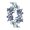
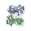
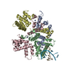
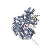
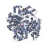
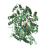
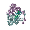
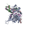
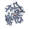
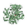
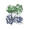
F: transfer RNA A: tRNA pseudouridine synthase A B: tRNA pseudouridine synthase A C: tRNA pseudouridine synthase A D: tRNA pseudouridine synthase A hetero molecules
Molecules of id A, B form a biological assembly unit of dimeric TruA. Molecules of id C, D and F form a biological assembly unit (C and D form a dimeric TruA enzyme and F is the substrate tRNA bound to the enzyme).
-
Components
#1: RNA chain
transferRNA
Mass: 28075.693 Da / Num. of mol.: 1 / Source method: obtained synthetically Details: Occurs naturally in E. coli. tRNA is obtained by in vitro transcription. References: GenBank: 174437
#2: Protein
tRNApseudouridinesynthaseA / tRNA-uridine isomerase I / tRNA pseudouridylate synthase I / PSU-I
Mass: 30441.615 Da / Num. of mol.: 4 Source method: isolated from a genetically manipulated source Source: (gene. exp.) Escherichia coli K12 (bacteria) / Species: Escherichia coli / Strain: K-12 / Gene: truA / Plasmid: pET28b / Species (production host): Escherichia coli / Production host: Escherichia coli BL21(DE3) (bacteria) / Strain (production host): BL21/DE3 References: UniProt: P07649, tRNA pseudouridine38-40 synthase
Resolution: 3.5→3.63 Å / Redundancy: 5.8 % / Mean I/σ(I) obs: 2.42 / Rsym value: 0.722 / % possible all: 99.9
-
Processing
Software
Name
Version
Classification
REFMAC
5.2.0019
refinement
ADSC
QUANTUM
datacollection
DENZO
datareduction
SCALEPACK
datascaling
PHASER
phasing
Refinement
Method to determine structure: MOLECULAR REPLACEMENT / Resolution: 3.5→20 Å / Cor.coef. Fo:Fc: 0.837 / Cor.coef. Fo:Fc free: 0.797 / SU B: 31.577 / SU ML: 0.501 / Cross valid method: THROUGHOUT / ESU R Free: 0.694 / Stereochemistry target values: MAXIMUM LIKELIHOOD / Details: HYDROGENS HAVE BEEN ADDED IN THE RIDING POSITIONS
Rfactor
Num. reflection
% reflection
Selection details
Rfree
0.27912
2384
10 %
RANDOM
Rwork
0.24601
-
-
-
obs
0.24936
21541
94.09 %
-
all
-
21541
-
-
Solvent computation
Ion probe radii: 0.8 Å / Shrinkage radii: 0.8 Å / VDW probe radii: 1.4 Å / Solvent model: BABINET MODEL WITH MASK
Displacement parameters
Biso mean: 37.066 Å2
Baniso -1
Baniso -2
Baniso -3
1-
-0.79 Å2
0 Å2
0 Å2
2-
-
-0.34 Å2
0 Å2
3-
-
-
1.13 Å2
Refinement step
Cycle: LAST / Resolution: 3.5→20 Å
Protein
Nucleic acid
Ligand
Solvent
Total
Num. atoms
8318
1441
6
0
9765
Refine LS restraints
Refine-ID
Type
Dev ideal
Dev ideal target
Number
X-RAY DIFFRACTION
r_bond_refined_d
0.01
0.021
10103
X-RAY DIFFRACTION
r_bond_other_d
X-RAY DIFFRACTION
r_angle_refined_deg
1.241
2.118
14080
X-RAY DIFFRACTION
r_angle_other_deg
X-RAY DIFFRACTION
r_dihedral_angle_1_deg
1.046
5
1050
X-RAY DIFFRACTION
r_dihedral_angle_2_deg
37.68
22.592
409
X-RAY DIFFRACTION
r_dihedral_angle_3_deg
16.62
15
1295
X-RAY DIFFRACTION
r_dihedral_angle_4_deg
17.399
15
78
X-RAY DIFFRACTION
r_chiral_restr
0.074
0.2
1602
X-RAY DIFFRACTION
r_gen_planes_refined
0.009
0.02
7325
X-RAY DIFFRACTION
r_gen_planes_other
X-RAY DIFFRACTION
r_nbd_refined
0.277
0.2
5149
X-RAY DIFFRACTION
r_nbd_other
X-RAY DIFFRACTION
r_nbtor_refined
0.335
0.2
6923
X-RAY DIFFRACTION
r_nbtor_other
X-RAY DIFFRACTION
r_xyhbond_nbd_refined
0.176
0.2
361
X-RAY DIFFRACTION
r_xyhbond_nbd_other
X-RAY DIFFRACTION
r_metal_ion_refined
0.14
0.2
6
X-RAY DIFFRACTION
r_metal_ion_other
X-RAY DIFFRACTION
r_symmetry_vdw_refined
0.253
0.2
56
X-RAY DIFFRACTION
r_symmetry_vdw_other
X-RAY DIFFRACTION
r_symmetry_hbond_refined
0.045
0.2
1
X-RAY DIFFRACTION
r_symmetry_hbond_other
X-RAY DIFFRACTION
r_symmetry_metal_ion_refined
X-RAY DIFFRACTION
r_symmetry_metal_ion_other
X-RAY DIFFRACTION
r_mcbond_it
1.315
1.5
5265
X-RAY DIFFRACTION
r_mcbond_other
X-RAY DIFFRACTION
r_mcangle_it
2.428
2
8473
X-RAY DIFFRACTION
r_scbond_it
1.653
3
4838
X-RAY DIFFRACTION
r_scangle_it
2.697
4.5
5607
X-RAY DIFFRACTION
r_rigid_bond_restr
X-RAY DIFFRACTION
r_sphericity_free
X-RAY DIFFRACTION
r_sphericity_bonded
LS refinement shell
Resolution: 3.5→3.588 Å / Total num. of bins used: 20
Rfactor
Num. reflection
% reflection
Rfree
0.283
178
-
Rwork
0.293
1404
-
obs
-
-
86.97 %
Refinement TLS params.
Method: refined / Refine-ID: X-RAY DIFFRACTION
ID
L11 (°2)
L12 (°2)
L13 (°2)
L22 (°2)
L23 (°2)
L33 (°2)
S11 (Å °)
S12 (Å °)
S13 (Å °)
S21 (Å °)
S22 (Å °)
S23 (Å °)
S31 (Å °)
S32 (Å °)
S33 (Å °)
T11 (Å2)
T12 (Å2)
T13 (Å2)
T22 (Å2)
T23 (Å2)
T33 (Å2)
Origin x (Å)
Origin y (Å)
Origin z (Å)
1
9.4025
4.3001
-4.3992
1.9666
-2.0119
2.0583
2.3087
-3.4701
2.4214
-1.3341
-1.0997
-0.0339
-0.2112
2.6433
-1.209
-0.6842
-0.2431
-0.0511
0.8427
-0.7294
1.1413
-1.7309
-40.8644
16.8144
2
5.5071
5.1265
2.1604
5.9912
3.6837
3.1427
0.5471
0.0867
0.8314
-0.0318
0.3566
-0.1557
0.0705
-0.0678
-0.9037
-0.0978
-0.0346
0.0316
-0.1194
-0.0555
0.0454
-22.4499
-38.7384
19.6135
3
9.4585
0.7976
2.9923
0.6179
0.3845
0.9784
0.2521
-0.2696
0.1278
0.256
0.0903
-0.2091
0.3509
-0.5243
-0.3423
-0.3149
-0.1055
0.0928
0.0047
-0.0619
0.1563
-35.8109
-25.6494
26.8037
4
17.3805
16.2064
-8.689
15.1117
-8.1021
4.3439
0.1815
3.22
0.5861
-0.6099
0.4868
4.709
-2.8809
-1.6243
-0.6682
1.3192
0.5303
0.1461
0.4724
0.1714
0.5749
-23.6234
-26.3229
5.4124
5
8.8017
2.5832
-3.4726
6.6673
6.1862
10.156
0.0368
0.5358
0.4361
0.0243
0.8224
-0.3217
0.5389
0.741
-0.8592
-0.2495
-0.0164
-0.0659
-0.0624
-0.1617
0.0471
-11.8783
-43.8419
2.7412
Refinement TLS group
ID
Refine-ID
Refine TLS-ID
Auth asym-ID
Label asym-ID
Auth seq-ID
Label seq-ID
1
X-RAY DIFFRACTION
1
F
A
3 - 7
3 - 7
2
X-RAY DIFFRACTION
1
F
A
66 - 70
77 - 81
3
X-RAY DIFFRACTION
2
F
A
8 - 15
8 - 15
4
X-RAY DIFFRACTION
2
F
A
20 - 25
20 - 26
5
X-RAY DIFFRACTION
3
F
A
26 - 47
27 - 58
6
X-RAY DIFFRACTION
5
F
A
48 - 65
59 - 76
7
X-RAY DIFFRACTION
5
F
A
16 - 20
16 - 20
+
About Yorodumi
-
News
-
Feb 9, 2022. New format data for meta-information of EMDB entries
New format data for meta-information of EMDB entries
Version 3 of the EMDB header file is now the official format.
The previous official version 1.9 will be removed from the archive.
In the structure databanks used in Yorodumi, some data are registered as the other names, "COVID-19 virus" and "2019-nCoV". Here are the details of the virus and the list of structure data.
Jan 31, 2019. EMDB accession codes are about to change! (news from PDBe EMDB page)
EMDB accession codes are about to change! (news from PDBe EMDB page)
The allocation of 4 digits for EMDB accession codes will soon come to an end. Whilst these codes will remain in use, new EMDB accession codes will include an additional digit and will expand incrementally as the available range of codes is exhausted. The current 4-digit format prefixed with “EMD-” (i.e. EMD-XXXX) will advance to a 5-digit format (i.e. EMD-XXXXX), and so on. It is currently estimated that the 4-digit codes will be depleted around Spring 2019, at which point the 5-digit format will come into force.
The EM Navigator/Yorodumi systems omit the EMD- prefix.
Related info.:Q: What is EMD? / ID/Accession-code notation in Yorodumi/EM Navigator
Yorodumi is a browser for structure data from EMDB, PDB, SASBDB, etc.
This page is also the successor to EM Navigator detail page, and also detail information page/front-end page for Omokage search.
The word "yorodu" (or yorozu) is an old Japanese word meaning "ten thousand". "mi" (miru) is to see.
Related info.:EMDB / PDB / SASBDB / Comparison of 3 databanks / Yorodumi Search / Aug 31, 2016. New EM Navigator & Yorodumi / Yorodumi Papers / Jmol/JSmol / Function and homology information / Changes in new EM Navigator and Yorodumi
 Movie
Movie Controller

 Yorodumi
 Open data
 Basic information
 Components
 Keywords
 Function and homology information
 X-RAY DIFFRACTION /
  Authors
 Citation
 Structure visualization
 Downloads & links
 Other downloads
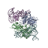


 PDBj
PDBj


 Assembly
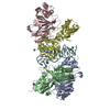


 Mass: 39.098 Da / Num. of mol.: 6 / Source method: obtained synthetically / Formula: K
 Sample preparation
 / Beamline: 8.3.1 / Wavelength: 1 Å
 Processing