[English] 日本語
Yorodumi
- PDB-2mwo: Solution structure of 53BP1 tandem Tudor domains in complex with ... -

+
Open data


ID or keywords:

Loading...

-
Basic information

Entry
Database: PDB / ID: 2mwo
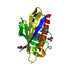
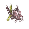
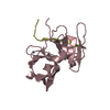
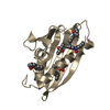
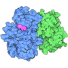
TitleSolution structure of 53BP1 tandem Tudor domains in complex with a p53K370me2 peptide
Components
  • Cellular tumor antigen p53
  • Tumor suppressor p53-binding protein 1
KeywordsTRANSCRIPTION/ANTITUMOR PROTEIN / Cell cycle / DNA damage response / TRANSCRIPTION-ANTITUMOR PROTEIN complex
Function / homology
Function and homology information


histone H4K20me methyltransferase activity / positive regulation of isotype switching / ubiquitin-modified histone reader activity / histone H4K20me2 reader activity / double-strand break repair via classical nonhomologous end joining / protein localization to site of double-strand break / negative regulation of helicase activity / Loss of function of TP53 in cancer due to loss of tetramerization ability / Regulation of TP53 Expression / signal transduction by p53 class mediator ...histone H4K20me methyltransferase activity / positive regulation of isotype switching / ubiquitin-modified histone reader activity / histone H4K20me2 reader activity / double-strand break repair via classical nonhomologous end joining / protein localization to site of double-strand break / negative regulation of helicase activity / Loss of function of TP53 in cancer due to loss of tetramerization ability / Regulation of TP53 Expression / signal transduction by p53 class mediator / negative regulation of G1 to G0 transition / Transcriptional activation of cell cycle inhibitor p21 / negative regulation of pentose-phosphate shunt / Activation of NOXA and translocation to mitochondria / ATP-dependent DNA/DNA annealing activity / regulation of cell cycle G2/M phase transition / DNA repair complex / oligodendrocyte apoptotic process / positive regulation of thymocyte apoptotic process / oxidative stress-induced premature senescence / bone marrow development / circadian behavior / cellular response to actinomycin D / positive regulation of programmed necrotic cell death / histone deacetylase regulator activity / RUNX3 regulates CDKN1A transcription / TP53 Regulates Transcription of Death Receptors and Ligands / Activation of PUMA and translocation to mitochondria / TP53 regulates transcription of additional cell cycle genes whose exact role in the p53 pathway remain uncertain / mRNA transcription / Urea cycle / Regulation of TP53 Activity through Association with Co-factors / ER overload response / hematopoietic stem cell differentiation / Formation of Senescence-Associated Heterochromatin Foci (SAHF) / TP53 Regulates Transcription of Caspase Activators and Caspases / intrinsic apoptotic signaling pathway by p53 class mediator / entrainment of circadian clock by photoperiod / Zygotic genome activation (ZGA) / telomeric repeat DNA binding / TP53 Regulates Transcription of Genes Involved in Cytochrome C Release / PI5P Regulates TP53 Acetylation / positive regulation of release of cytochrome c from mitochondria / hematopoietic progenitor cell differentiation / Association of TriC/CCT with target proteins during biosynthesis / negative regulation of telomere maintenance via telomerase / SUMOylation of transcription factors / TP53 regulates transcription of several additional cell death genes whose specific roles in p53-dependent apoptosis remain uncertain / intrinsic apoptotic signaling pathway in response to DNA damage by p53 class mediator / Transcriptional Regulation by VENTX / replicative senescence / TFIID-class transcription factor complex binding / intrinsic apoptotic signaling pathway in response to endoplasmic reticulum stress / viral process / positive regulation of intrinsic apoptotic signaling pathway / Pyroptosis / determination of adult lifespan / positive regulation of RNA polymerase II transcription preinitiation complex assembly / negative regulation of fibroblast proliferation / general transcription initiation factor binding / positive regulation of execution phase of apoptosis / type II interferon-mediated signaling pathway / TP53 Regulates Transcription of Genes Involved in G1 Cell Cycle Arrest / negative regulation of double-strand break repair via homologous recombination / positive regulation of intrinsic apoptotic signaling pathway by p53 class mediator / cellular response to glucose starvation / core promoter sequence-specific DNA binding / cis-regulatory region sequence-specific DNA binding / Regulation of TP53 Activity through Acetylation / DNA damage checkpoint signaling / intrinsic apoptotic signaling pathway / mitotic G1 DNA damage checkpoint signaling / histone reader activity / transcription repressor complex / response to gamma radiation / tumor necrosis factor-mediated signaling pathway / 14-3-3 protein binding / MDM2/MDM4 family protein binding / TP53 Regulates Transcription of Genes Involved in G2 Cell Cycle Arrest / protein phosphatase 2A binding / molecular function activator activity / transcription initiation-coupled chromatin remodeling / Regulation of PTEN gene transcription / cellular response to ionizing radiation / cellular response to xenobiotic stimulus / DNA damage response, signal transduction by p53 class mediator / TP53 Regulates Metabolic Genes / double-strand break repair via nonhomologous end joining / TP53 Regulates Transcription of DNA Repair Genes / replication fork / Regulation of NF-kappa B signaling / mRNA 3'-UTR binding / protein tetramerization / autophagy / promoter-specific chromatin binding / Nonhomologous End-Joining (NHEJ) / Stabilization of p53 / molecular condensate scaffold activity / negative regulation of cell growth / cellular response to gamma radiation
Similarity search - Function
Tumour suppressor p53-binding protein-1 Tudor domain / : / Tumour suppressor p53-binding protein-1 Tudor / BRCA1 C Terminus (BRCT) domain / : / : / SH3 type barrels. - #30 / Cellular tumor antigen p53, transactivation domain 2 / Transactivation domain 2 / p53 transactivation domain ...Tumour suppressor p53-binding protein-1 Tudor domain / : / Tumour suppressor p53-binding protein-1 Tudor / BRCA1 C Terminus (BRCT) domain / : / : / SH3 type barrels. - #30 / Cellular tumor antigen p53, transactivation domain 2 / Transactivation domain 2 / p53 transactivation domain / P53 transactivation motif / SH3 type barrels. - #140 / : / p53 family signature. / p53, tetramerisation domain / P53 tetramerisation motif / p53, DNA-binding domain / P53 DNA-binding domain / p53 tumour suppressor family / p53-like tetramerisation domain superfamily / p53/RUNT-type transcription factor, DNA-binding domain superfamily / p53-like transcription factor, DNA-binding / breast cancer carboxy-terminal domain / BRCT domain profile. / BRCT domain / BRCT domain superfamily / SH3 type barrels. / Roll / Ribosomal protein L2, domain 2 / Mainly Beta
Similarity search - Domain/homology
Cellular tumor antigen p53 / TP53-binding protein 1
Similarity search - Component
Biological speciesHomo sapiens (human)
MethodSOLUTION NMR / simulated annealing
Model detailslowest energy, model1
AuthorsCui, G. / Botuyan, M.V. / Mer, G.
CitationJournal: Structure / Year: 2015
Title: Structural Plasticity of Methyllysine Recognition by the Tandem Tudor Domain of 53BP1.
Authors: Tong, Q. / Cui, G. / Botuyan, M.V. / Rothbart, S.B. / Hayashi, R. / Musselman, C.A. / Singh, N. / Appella, E. / Strahl, B.D. / Mer, G. / Kutateladze, T.G.
History
DepositionNov 15, 2014Deposition site: BMRB / Processing site: RCSB
Revision 1.0Dec 10, 2014Provider: repository / Type: Initial release
Revision 1.1Jan 21, 2015Group: Database references
Revision 1.2Mar 18, 2015Group: Database references
Revision 1.3Mar 26, 2025Group: Data collection / Database references ...Data collection / Database references / Derived calculations / Structure summary
Category: chem_comp_atom / chem_comp_bond ...chem_comp_atom / chem_comp_bond / database_2 / pdbx_entry_details / pdbx_modification_feature / pdbx_nmr_software / pdbx_nmr_spectrometer / struct_conn / struct_ref_seq_dif / struct_site
Item: _database_2.pdbx_DOI / _database_2.pdbx_database_accession ..._database_2.pdbx_DOI / _database_2.pdbx_database_accession / _pdbx_nmr_software.name / _pdbx_nmr_spectrometer.model / _struct_conn.pdbx_leaving_atom_flag / _struct_ref_seq_dif.details / _struct_site.pdbx_auth_asym_id / _struct_site.pdbx_auth_comp_id / _struct_site.pdbx_auth_seq_id

-
Structure visualization

Structure viewerMolecule:
MolmilJmol/JSmol

Downloads & links

-
Assembly

Deposited unit
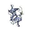
A: Tumor suppressor p53-binding protein 1
B: Cellular tumor antigen p53


Theoretical massNumber of molelcules
Total (without water)15,6302
Polymers15,6302
Non-polymers00
Water00
1


  • Idetical with deposited unit
  • defined by author&software
TypeNameSymmetry operationNumber
identity operation1_555x,y,z1
Buried area690.9 Å2
ΔGint-9.2 kcal/mol
Surface area2060 Å2
MethodPISA
NMR ensembles
DataCriteria
Number of conformers (submitted / calculated)20 / 200structures with the lowest energy
RepresentativeModel #1lowest energy

-
Components

#1: Protein Tumor suppressor p53-binding protein 1 / 53BP1 / p53-binding protein 1 / p53BP1


Mass: 13944.780 Da / Num. of mol.: 1 / Fragment: Tudor-like region residues 1484-1603
Source method: isolated from a genetically manipulated source
Source: (gene. exp.) Homo sapiens (human) / Gene: TP53BP1 / Production host: Escherichia coli (E. coli) / References: UniProt: Q12888
#2: Protein/peptide Cellular tumor antigen p53 / p53K370Me2 / Antigen NY-CO-13 / Phosphoprotein p53 / Tumor suppressor p53


Mass: 1684.919 Da / Num. of mol.: 1 / Fragment: DNA-binding repression region residues 363-377 / Source method: obtained synthetically / Source: (synth.) Homo sapiens (human) / References: UniProt: P04637
Has protein modificationY

-
Experimental details

-
Experiment

ExperimentMethod: SOLUTION NMR
NMR experiment
Conditions-IDExperiment-IDSolution-IDType
1112D 1H-15N HSQC
1232D 1H-15N HSQC
1322D 1H-13C HSQC
1442D 1H-13C HSQC
1522D 1H-13C HSQC aromatic
1613D HNCA
1713D HN(CA)CB
1833D HN(CA)CB
1913D HNCO
11013D CBCA(CO)NH
11113D HN(CA)CO
11213D H(CCO)NH
11313D C(CO)NH
11413D HBHA(CO)NH
11513D (H)CCH-TOCSY
11652D 1H-15N HSQC
11753D 1H-15N TOCSY
11823D 1H-13C NOESY aliphatic
11923D 1H-13C NOESY aromatic
12053D 1H-15N NOESY
12123D 1H-15N NOESY
12223D Filtered (15N/13C)-Edited (13C)
12343D Filtered (15N/13C)-Edited (13C)
12423D (HB)CB(CGCD)HD
12562D 1H-13C HSQC
12672D 1H-13C HSQC
12772D 1H-15N HSQC

-
Sample preparation

Details
Solution-IDContentsSolvent system
125 mM sodium phosphate, 1.5 mM sodium azide, 2.0 mM [U-100% 13C; U-100% 15N] 53BP1-Tudor, 6.0 mM p53K370me2, 90% H2O/10% D2O90% H2O/10% D2O
225 mM sodium phosphate, 1.5 mM sodium azide, 2.0 mM [U-100% 13C; U-100% 15N] 53BP1-Tudor, 6.0 mM p53K370me2, 100% D2O100% D2O
325 mM sodium phosphate, 1.5 mM sodium azide, 5.0 mM 53BP1-Tudor, 2.0 mM [U-100% 13C; U-100% 15N] p53Kc370me2, 90% H2O/10% D2O90% H2O/10% D2O
425 mM sodium phosphate, 1.5 mM sodium azide, 5.0 mM 53BP1-Tudor, 2.0 mM [U-100% 13C; U-100% 15N] p53Kc370me2, 100% D2O100% D2O
525 mM sodium phosphate, 1.5 mM sodium azide, 2.0 mM [U-100% 15N] 53BP1-Tudor, 6.0 mM p53K370me2, 90% H2O/10% D2O90% H2O/10% D2O
635 mM sodium phosphate, 1.5 mM sodium azide, 2.0 mM p53K370me2, 100% D2O100% D2O
725 mM sodium phosphate, 1.5 mM sodium azide, 2.0 mM [U-100% 13C; U-100% 15N] p53Kc370me2, 100% D2O100% D2O
Sample
Conc. (mg/ml)ComponentIsotopic labelingSolution-ID
25 mMsodium phosphate-11
1.5 mMsodium azide-21
2.0 mM53BP1-Tudor-3[U-100% 13C; U-100% 15N]1
6.0 mMp53K370me2-41
25 mMsodium phosphate-52
1.5 mMsodium azide-62
2.0 mM53BP1-Tudor-7[U-100% 13C; U-100% 15N]2
6.0 mMp53K370me2-82
25 mMsodium phosphate-93
1.5 mMsodium azide-103
5.0 mM53BP1-Tudor-113
2.0 mMp53Kc370me2-12[U-100% 13C; U-100% 15N]3
25 mMsodium phosphate-134
1.5 mMsodium azide-144
5.0 mM53BP1-Tudor-154
2.0 mMp53Kc370me2-16[U-100% 13C; U-100% 15N]4
25 mMsodium phosphate-175
1.5 mMsodium azide-185
2.0 mM53BP1-Tudor-19[U-100% 15N]5
6.0 mMp53K370me2-205
35 mMsodium phosphate-216
1.5 mMsodium azide-226
2.0 mMp53K370me2-236
25 mMsodium phosphate-247
1.5 mMsodium azide-257
2.0 mMp53Kc370me2-26[U-100% 13C; U-100% 15N]7
Sample conditionsIonic strength: 25 / pH: 7.5 / Pressure: ambient / Temperature: 298 K

-
NMR measurement

NMR spectrometerType: Bruker Avance III / Manufacturer: Bruker / Model: AVANCE III / Field strength: 700 MHz

-
Processing

NMR software
NameDeveloperClassification
XwinNMRBruker Biospincollection
NMRPipeDelaglio, Grzesiek, Vuister, Zhu, Pfeifer and Baxprocessing
NMRViewJohnson, One Moon Scientificprocessing
SANEDuggan, Legge, Dyson & Wrightchemical shift assignment
TALOSCornilescu, Delaglio and Baxdata analysis
CYANAGuntert, Mumenthaler and Wuthrichstructure solution
AmberCase, Darden, Cheatham, III, Simmerling, Wang, Duke, Luo, and Kollmanrefinement
RefinementMethod: simulated annealing / Software ordinal: 1
NMR representativeSelection criteria: lowest energy
NMR ensembleConformer selection criteria: structures with the lowest energy
Conformers calculated total number: 200 / Conformers submitted total number: 20

+
About Yorodumi

-
News

-
Feb 9, 2022. New format data for meta-information of EMDB entries

New format data for meta-information of EMDB entries

  • Version 3 of the EMDB header file is now the official format.
  • The previous official version 1.9 will be removed from the archive.

Related info.:EMDB header

External links:wwPDB to switch to version 3 of the EMDB data model

-
Aug 12, 2020. Covid-19 info

Covid-19 info

URL: https://pdbj.org/emnavi/covid19.php

New page: Covid-19 featured information page in EM Navigator.

Related info.:Covid-19 info / Mar 5, 2020. Novel coronavirus structure data

+
Mar 5, 2020. Novel coronavirus structure data

Novel coronavirus structure data

Related info.:Yorodumi Speices / Aug 12, 2020. Covid-19 info

External links:COVID-19 featured content - PDBj / Molecule of the Month (242):Coronavirus Proteases

+
Jan 31, 2019. EMDB accession codes are about to change! (news from PDBe EMDB page)

EMDB accession codes are about to change! (news from PDBe EMDB page)

  • The allocation of 4 digits for EMDB accession codes will soon come to an end. Whilst these codes will remain in use, new EMDB accession codes will include an additional digit and will expand incrementally as the available range of codes is exhausted. The current 4-digit format prefixed with “EMD-” (i.e. EMD-XXXX) will advance to a 5-digit format (i.e. EMD-XXXXX), and so on. It is currently estimated that the 4-digit codes will be depleted around Spring 2019, at which point the 5-digit format will come into force.
  • The EM Navigator/Yorodumi systems omit the EMD- prefix.

Related info.:Q: What is EMD? / ID/Accession-code notation in Yorodumi/EM Navigator

External links:EMDB Accession Codes are Changing Soon! / Contact to PDBj

+
Jul 12, 2017. Major update of PDB

Major update of PDB

  • wwPDB released updated PDB data conforming to the new PDBx/mmCIF dictionary.
  • This is a major update changing the version number from 4 to 5, and with Remediation, in which all the entries are updated.
  • In this update, many items about electron microscopy experimental information are reorganized (e.g. em_software).
  • Now, EM Navigator and Yorodumi are based on the updated data.

External links:wwPDB Remediation / Enriched Model Files Conforming to OneDep Data Standards Now Available in the PDB FTP Archive

-
Yorodumi

Thousand views of thousand structures

  • Yorodumi is a browser for structure data from EMDB, PDB, SASBDB, etc.
  • This page is also the successor to EM Navigator detail page, and also detail information page/front-end page for Omokage search.
  • The word "yorodu" (or yorozu) is an old Japanese word meaning "ten thousand". "mi" (miru) is to see.

Related info.:EMDB / PDB / SASBDB / Comparison of 3 databanks / Yorodumi Search / Aug 31, 2016. New EM Navigator & Yorodumi / Yorodumi Papers / Jmol/JSmol / Function and homology information / Changes in new EM Navigator and Yorodumi

Read more