 Movie
Movie Controller

[English] 日本語
 Yorodumi
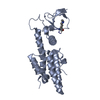
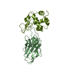
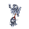
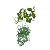
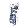
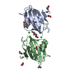
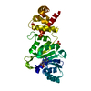
- PDB-2msb: STRUCTURE OF A C-TYPE MANNOSE-BINDING PROTEIN COMPLEXED WITH AN O... -
+ Open data

- Basic information
| Entry | Database: PDB / ID: 2msb | |||||||||
|---|---|---|---|---|---|---|---|---|---|---|
| Title | STRUCTURE OF A C-TYPE MANNOSE-BINDING PROTEIN COMPLEXED WITH AN OLIGOSACCHARIDE | |||||||||
 Components Components | MANNOSE-BINDING PROTEIN-A | |||||||||
 Keywords Keywords | LECTIN | |||||||||
| Function / homology |  Function and homology information Function and homology informationcalcium-dependent carbohydrate binding / complement activation, lectin pathway / : / oligosaccharide binding / collagen trimer / surfactant homeostasis / phosphatidylinositol-4-phosphate binding / protein homotrimerization / D-mannose binding / polysaccharide binding ...calcium-dependent carbohydrate binding / complement activation, lectin pathway / : / oligosaccharide binding / collagen trimer / surfactant homeostasis / phosphatidylinositol-4-phosphate binding / protein homotrimerization / D-mannose binding / polysaccharide binding / complement activation, classical pathway / multivesicular body / positive regulation of phagocytosis / calcium-dependent protein binding / protease binding / defense response to Gram-positive bacterium / calcium ion binding / protein homodimerization activity / : / identical protein binding Similarity search - Function | |||||||||
| Biological species |  | |||||||||
| Method |  X-RAY DIFFRACTION / Resolution: 1.7 Å X-RAY DIFFRACTION / Resolution: 1.7 Å | |||||||||
 Authors Authors | Weis, W.I. / Drickamer, K. / Hendrickson, W.A. | |||||||||
 Citation Citation | Journal: Nature / Year: 1992 Title: Structure of a C-type mannose-binding protein complexed with an oligosaccharide. Authors: Weis, W.I. / Drickamer, K. / Hendrickson, W.A. #1: Journal: J.Biol.Chem. / Year: 1991Title: Physical Characterization and Crystallization of the Carbohydrate-Recognition Domain of a Mannose-Binding Protein from Rat Authors: Weis, W.I. / Crichlow, G.V. / Murthy, H.M.K. / Hendrickson, W.A. / Drickamer, K. #2: Journal: Science / Year: 1991Title: Structure of the Calcium-Dependent Lectin Domain from a Rat Mannose-Binding Protein Determined by MAD Phasing Authors: Weis, W.I. / Kahn, R. / Fourme, R. / Drickamer, K. / Hendrickson, W.A. | |||||||||
| History |
|
- Structure visualization
| Structure viewer | Molecule: MolmilJmol/JSmol |
|---|
- Downloads & links
-Download
| PDBx/mmCIF format | 2msb.cif.gz | 68.1 KB | Display | PDBx/mmCIF format |
|---|---|---|---|---|
| PDB format | pdb2msb.ent.gz | 48.1 KB | Display | PDB format |
| PDBx/mmJSON format | 2msb.json.gz | Tree view | PDBx/mmJSON format | |
| Others |  Other downloads Other downloads |
-Validation report
| Arichive directory | https://data.pdbj.org/pub/pdb/validation_reports/ms/2msbftp://data.pdbj.org/pub/pdb/validation_reports/ms/2msb | HTTPS FTP |
|---|
-Related structure data
| Similar structure data |
|---|


-Links
 PDBj
PDBj


- Assembly
| Deposited unit | 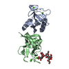
| ||||||||
|---|---|---|---|---|---|---|---|---|---|
| 1 |
| ||||||||
| Unit cell |
| ||||||||
| Atom site foot note | 1: RESIDUES PRO A 186 AND PRO B 186 ARE CIS PROLINES. | ||||||||
| Noncrystallographic symmetry (NCS) | NCS oper: (Code: given Matrix: (0.21747, -0.34809, -0.91189), Vector: Details | THE ASYMMETRIC UNIT OF THE CRYSTAL CONTAINS ONE DIMERIC PROTEIN MOLECULE (2 IDENTICAL CHAINS CORRESPONDING TO RESIDUES 107 - 221 MBP-A), AND ONE GLYCOPEPTIDE MOLECULE (6 MANNOSE, 2 GLCNAC (NAG), 1 ASN). FIVE OF THE MANNOSE RESIDUES CROSSLINK NEIGHBORING DIMERS OF THE CRYSTAL. MAN 9 BINDS TO CHAIN 2, AND MAN 8 BINDS TO THE (-X, Y + 1/2, 1 - Z) SYMMETRY MATE OF CHAIN 1. THE NON-CRYSTALLOGRAPHIC SYMMETRY TRANSFORMATION PRESENTED ON *MTRIX* RECORDS BELOW WILL YIELD APPROXIMATE COORDINATES FOR CHAIN *A* WHEN APPLIED TO CHAIN *B*. THE TRANSFORMATION WAS DERIVED BY LEAST-SQUARES SUPERPOSITION OF THE MAIN CHAIN N, CA, C, O AND CB, WHERE PRESENT, OF RESIDUES 110 - 220. | |
-Components
| #1: Protein | Mass: 12688.150 Da / Num. of mol.: 2 Source method: isolated from a genetically manipulated source Source: (gene. exp.) #2: Polysaccharide | alpha-D-mannopyranose-(1-2)-alpha-D-mannopyranose-(1-3)-[alpha-D-mannopyranose-(1-3)-[alpha-D- ...alpha-D-mannopyranose-(1-2)-alpha-D-mannopyranose-(1-3)-[alpha-D-mannopyranose-(1-3)-[alpha-D-mannopyranose-(1-6)]alpha-D-mannopyranose-(1-6)]beta-D-mannopyranose-(1-4)-2-acetamido-2-deoxy-beta-D-glucopyranose | Source method: isolated from a genetically manipulated source #3: Chemical | ChemComp-CA /   Mass: 40.078 Da / Num. of mol.: 6 / Source method: obtained synthetically / Formula: Ca Mass: 40.078 Da / Num. of mol.: 6 / Source method: obtained synthetically / Formula: Ca#4: Water | ChemComp-HOH / |  Mass: 18.015 Da / Num. of mol.: 211 / Source method: isolated from a natural source / Formula: H2O Mass: 18.015 Da / Num. of mol.: 211 / Source method: isolated from a natural source / Formula: H2OHas protein modification | Y | |
|---|
-Experimental details
-Experiment
| Experiment | Method: X-RAY DIFFRACTION |
|---|
- Sample preparation
| Crystal | Density Matthews: 2.29 Å3/Da / Density % sol: 46.29 % | ||||||||||||||||||||||||||||||||||||||||||||||||||||||||||||||||||||||
|---|---|---|---|---|---|---|---|---|---|---|---|---|---|---|---|---|---|---|---|---|---|---|---|---|---|---|---|---|---|---|---|---|---|---|---|---|---|---|---|---|---|---|---|---|---|---|---|---|---|---|---|---|---|---|---|---|---|---|---|---|---|---|---|---|---|---|---|---|---|---|---|
| Crystal grow | *PLUS pH: 8 / Method: unknown | ||||||||||||||||||||||||||||||||||||||||||||||||||||||||||||||||||||||
| Components of the solutions | *PLUS
|
-Data collection
| Reflection | *PLUS Highest resolution: 1.7 Å / Lowest resolution: 10 Å / Num. obs: 22750 / % possible obs: 91 % / Observed criterion σ(I): 3 / Num. measured all: 17511 / Rmerge(I) obs: 0.07 |
|---|
- Processing
| Software |
| ||||||||||||||||||||||||||||||||||||||||||||||||||||||||||||
|---|---|---|---|---|---|---|---|---|---|---|---|---|---|---|---|---|---|---|---|---|---|---|---|---|---|---|---|---|---|---|---|---|---|---|---|---|---|---|---|---|---|---|---|---|---|---|---|---|---|---|---|---|---|---|---|---|---|---|---|---|---|
| Refinement | Resolution: 1.7→10 Å / σ(F): 3 Details: AN OVERALL ANISOTROPIC TEMPERATURE FACTOR IS REQUIRED TO CORRECTLY MODEL THE DATA, BUT HAS NOT BEEN APPLIED TO THE SUBMITTED MODEL (OR STRUCTURE FACTORS). THE VALUES OF THE TENSOR ARE: B(11) ...Details: AN OVERALL ANISOTROPIC TEMPERATURE FACTOR IS REQUIRED TO CORRECTLY MODEL THE DATA, BUT HAS NOT BEEN APPLIED TO THE SUBMITTED MODEL (OR STRUCTURE FACTORS). THE VALUES OF THE TENSOR ARE: B(11) = 4.1 ANGSTROMS**2 B(12) = B(23) = 0.0 ANGSTROMS**2 B(13) = 2.2 ANGSTROMS**2 B(22) = -4.0 ANGSTROMS**2 B(33) = -4.2 ANGSTROMS**2 THE TENSOR IS DEFINED IN SHERIFF, S. AND HENDRICKSON, W.A. (1987) ACTA CRYST. A43:118-121.
| ||||||||||||||||||||||||||||||||||||||||||||||||||||||||||||
| Refinement step | Cycle: LAST / Resolution: 1.7→10 Å
| ||||||||||||||||||||||||||||||||||||||||||||||||||||||||||||
| Refine LS restraints |
| ||||||||||||||||||||||||||||||||||||||||||||||||||||||||||||
| Refinement | *PLUS Highest resolution: 1.7 Å / Lowest resolution: 10 Å / Num. reflection obs: 20461 / σ(F): 3 / Rfactor obs: 0.174 | ||||||||||||||||||||||||||||||||||||||||||||||||||||||||||||
| Solvent computation | *PLUS | ||||||||||||||||||||||||||||||||||||||||||||||||||||||||||||
| Displacement parameters | *PLUS | ||||||||||||||||||||||||||||||||||||||||||||||||||||||||||||
| Refine LS restraints | *PLUS
|
