 Movie
Movie Controller


+ Open data

- Basic information
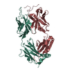
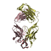
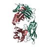
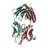
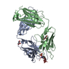
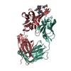
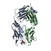
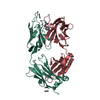
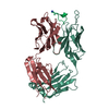
| Entry | Database: PDB / ID: 2mpa | |||||||||
|---|---|---|---|---|---|---|---|---|---|---|
| Title | BACTERICIDAL ANTIBODY AGAINST NEISSERIA MENINGITIDIS | |||||||||
 Components Components |
| |||||||||
 Keywords Keywords | IMMUNE SYSTEM / MURINE IMMUNOGLOBULIN IGG2A KAPPA / BACTERICIDAL ANTIBODY / EPITOPE P1.16 OF PORA FROM NEISSERIA MENINGITIDIS / COMPLEX (IMMUNOGLOBULIN-PEPTIDE) | |||||||||
| Function / homology |  Function and homology information Function and homology informationalpha-beta T cell receptor complex / IgG immunoglobulin complex / porin activity / pore complex / immunoglobulin mediated immune response / immunoglobulin complex / antigen binding / B cell differentiation / cell outer membrane / monoatomic ion transmembrane transport ...alpha-beta T cell receptor complex / IgG immunoglobulin complex / porin activity / pore complex / immunoglobulin mediated immune response / immunoglobulin complex / antigen binding / B cell differentiation / cell outer membrane / monoatomic ion transmembrane transport / adaptive immune response / extracellular region / metal ion binding / plasma membrane Similarity search - Function | |||||||||
| Biological species |  synthetic construct (others) | |||||||||
| Method |  X-RAY DIFFRACTION / MOLECULAR REPLACEMENT / Resolution: 2.6 Å X-RAY DIFFRACTION / MOLECULAR REPLACEMENT / Resolution: 2.6 Å | |||||||||
 Authors Authors | Van Den Elsen, J.M.H. / Herron, J.N. / Kroon, J. / Gros, P. | |||||||||
 Citation Citation | Journal: Proteins / Year: 1997 Title: Bactericidal antibody recognition of a PorA epitope of Neisseria meningitidis: crystal structure of a Fab fragment in complex with a fluorescein-conjugated peptide. Authors: van den Elsen, J.M. / Herron, J.N. / Hoogerhout, P. / Poolman, J.T. / Boel, E. / Logtenberg, T. / Wilting, J. / Crommelin, D.J. / Kroon, J. / Gros, P. #1: Journal: J.Biol.Chem. / Year: 1999Title: Bactericidal Antibody Recognition of Meningococcal Pora by Induced Fit: Comparison of Liganded and Unliganded Fab Structures Authors: van den Elsen, J. / Vandeputte-Rutten, L. / Kroon, J. / Gros, P. | |||||||||
| History |
|
- Structure visualization
| Structure viewer | Molecule: MolmilJmol/JSmol |
|---|
- Downloads & links
-Download
| PDBx/mmCIF format | 2mpa.cif.gz | 104.1 KB | Display | PDBx/mmCIF format |
|---|---|---|---|---|
| PDB format | pdb2mpa.ent.gz | 77.7 KB | Display | PDB format |
| PDBx/mmJSON format | 2mpa.json.gz | Tree view | PDBx/mmJSON format | |
| Others |  Other downloads Other downloads |
-Validation report
| Arichive directory | https://data.pdbj.org/pub/pdb/validation_reports/mp/2mpaftp://data.pdbj.org/pub/pdb/validation_reports/mp/2mpa | HTTPS FTP |
|---|
-Related structure data
| Related structure data | 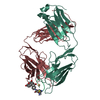 1mpaC  2hfl S: Starting model for refinement C: citing same article ( |
|---|---|
| Similar structure data |

-Links
 PDBj
PDBj


- Assembly
| Deposited unit | 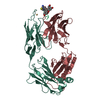
| ||||||||||
|---|---|---|---|---|---|---|---|---|---|---|---|
| 1 |
| ||||||||||
| Unit cell |
|
-Components
| #1: Antibody | Mass: 24122.861 Da / Num. of mol.: 1 / Fragment: FAB FRAGMENT / Source method: isolated from a natural source / Source: (natural) |
|---|---|
| #2: Antibody | Mass: 24415.350 Da / Num. of mol.: 1 / Fragment: FAB FRAGMENT / Source method: isolated from a natural source / Source: (natural) |
| #3: Protein/peptide | Mass: 1453.509 Da / Num. of mol.: 1 Fragment: APEX OF EXTRACELLULAR LOOP 4 (VR2) OF PORA, RESIDUES 180 - 187 Source method: obtained synthetically Details: SEQUENCE FROM NEISSERIA MENINGITIDIS (MENINGOCOCCUS), STRAIN: H44/76, VARIANT: P1.16; Source: (synth.) synthetic construct (others) / References: UniProt: E6MXW0*PLUS |
| #4: Chemical | ChemComp-CD /   Mass: 112.411 Da / Num. of mol.: 1 / Source method: obtained synthetically / Formula: Cd Mass: 112.411 Da / Num. of mol.: 1 / Source method: obtained synthetically / Formula: Cd |
| #5: Water | ChemComp-HOH /  Mass: 18.015 Da / Num. of mol.: 51 / Source method: isolated from a natural source / Formula: H2O Mass: 18.015 Da / Num. of mol.: 51 / Source method: isolated from a natural source / Formula: H2O |
| Nonpolymer details | CD: THIS PUTATIVE CADMIUM ION IS ASSIGNED TO A 6 SIGMA DENSITY PEAK IN A 2|FO|-|FC| MAP; IT IS ...CD: THIS PUTATIVE CADMIUM ION IS ASSIGNED TO A 6 SIGMA DENSITY PEAK IN A 2|FO|-|FC| MAP; IT IS COORDINATE |
| Sequence details | REFERENCE: THE MN12H2 LIGHT CHAIN AMINO ACID SEQUENCE IS DEDUCED FROM THE GENBANK U60442 NUCLEOTIDE ...REFERENCE: THE MN12H2 LIGHT CHAIN AMINO ACID SEQUENCE IS DEDUCED FROM THE GENBANK U60442 NUCLEOTIDE |
-Experimental details
-Experiment
| Experiment | Method: X-RAY DIFFRACTION / Number of used crystals: 1 |
|---|
- Sample preparation
| Crystal | Density Matthews: 2.6 Å3/Da / Density % sol: 53 % |
|---|---|
| Crystal grow | Method: vapor diffusion, hanging drop / pH: 4.6 Details: THE MN12H2 FAB PEPTIDE COMPLEX WAS CRYSTALLIZED FROM 15% W/V PEG 20000, 100 MM SODIUM ACETATE, PH 4.6, 20 MM CDCL2, VAPOR DIFFUSION, HANGING DROP |
| Crystal grow | *PLUS Method: unknown |
-Data collection
| Diffraction | Mean temperature: 298 K |
|---|---|
| Diffraction source | Source: ROTATING ANODE / Type: ENRAF-NONIUS FR571 / Wavelength: 1.5418 |
| Detector | Type: MAC Science DIP-2020 / Detector: IMAGE PLATE / Date: Mar 15, 1995 / Details: COLLIMATOR |
| Radiation | Monochromator: GRAPHITE / Protocol: SINGLE WAVELENGTH / Monochromatic (M) / Laue (L): M / Scattering type: x-ray |
| Radiation wavelength | Wavelength: 1.5418 Å / Relative weight: 1 |
| Reflection | Resolution: 2.6→20 Å / Num. obs: 15404 / % possible obs: 97 % / Observed criterion σ(I): 0 / Redundancy: 6.1 % / Rmerge(I) obs: 0.054 / Rsym value: 0.054 / Net I/σ(I): 16.8 |
| Reflection shell | Resolution: 2.6→2.64 Å / Redundancy: 2.5 % / Rmerge(I) obs: 0.357 / Mean I/σ(I) obs: 2.3 / Rsym value: 0.357 / % possible all: 96.5 |
- Processing
| Software |
| ||||||||||||||||||||||||||||||||||||||||||||||||||||||||||||||||||||||||||||||||
|---|---|---|---|---|---|---|---|---|---|---|---|---|---|---|---|---|---|---|---|---|---|---|---|---|---|---|---|---|---|---|---|---|---|---|---|---|---|---|---|---|---|---|---|---|---|---|---|---|---|---|---|---|---|---|---|---|---|---|---|---|---|---|---|---|---|---|---|---|---|---|---|---|---|---|---|---|---|---|---|---|---|
| Refinement | Method to determine structure: MOLECULAR REPLACEMENT Starting model: PDB ENTRY 2HFL 2hfl Resolution: 2.6→20 Å / Data cutoff high rms absF: 100000 / Isotropic thermal model: RESTRAINED / Cross valid method: THROUGHOUT / σ(F): 0
| ||||||||||||||||||||||||||||||||||||||||||||||||||||||||||||||||||||||||||||||||
| Solvent computation | Solvent model: FLAT MODEL / Bsol: 64.7 Å2 / ksol: 0.4 e/Å3 | ||||||||||||||||||||||||||||||||||||||||||||||||||||||||||||||||||||||||||||||||
| Displacement parameters | Biso mean: 47.6 Å2
| ||||||||||||||||||||||||||||||||||||||||||||||||||||||||||||||||||||||||||||||||
| Refine analyze |
| ||||||||||||||||||||||||||||||||||||||||||||||||||||||||||||||||||||||||||||||||
| Refinement step | Cycle: LAST / Resolution: 2.6→20 Å
| ||||||||||||||||||||||||||||||||||||||||||||||||||||||||||||||||||||||||||||||||
| Refine LS restraints |
| ||||||||||||||||||||||||||||||||||||||||||||||||||||||||||||||||||||||||||||||||
| LS refinement shell | Resolution: 2.6→2.69 Å / Total num. of bins used: 10 /
| ||||||||||||||||||||||||||||||||||||||||||||||||||||||||||||||||||||||||||||||||
| Xplor file |
|
