 Movie
Movie Controller

[English] 日本語
 Yorodumi
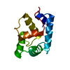
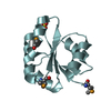
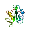
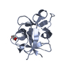
- PDB-2moi: 3D NMR structure of the cytoplasmic rhodanese domain of the inner... -
+ Open data

- Basic information
| Entry | Database: PDB / ID: 2moi | ||||||
|---|---|---|---|---|---|---|---|
| Title | 3D NMR structure of the cytoplasmic rhodanese domain of the inner membrane protein YgaP from Escherichia coli | ||||||
 Components Components | Inner membrane protein YgaP | ||||||
 Keywords Keywords | MEMBRANE PROTEIN / Rhodanese domain | ||||||
| Function / homology |  Function and homology information Function and homology informationthiosulfate sulfurtransferase / thiosulfate-cyanide sulfurtransferase activity / plasma membrane Similarity search - Function | ||||||
| Biological species |  | ||||||
| Method | SOLUTION NMR / simulated annealing | ||||||
| Model details | fewest violations, model 10 | ||||||
 Authors Authors | Eichmann, C. / Tzitzilonis, C. / Bordignon, E. / Maslennikov, I. / Choe, S. / Riek, R. | ||||||
 Citation Citation | Journal: J.Biol.Chem. / Year: 2014 Title: Solution NMR Structure and Functional Analysis of the Integral Membrane Protein YgaP from Escherichia coli. Authors: Eichmann, C. / Tzitzilonis, C. / Bordignon, E. / Maslennikov, I. / Choe, S. / Riek, R. | ||||||
| History |
|
- Structure visualization
| Structure viewer | Molecule: MolmilJmol/JSmol |
|---|
- Downloads & links
-Download
| PDBx/mmCIF format | 2moi.cif.gz | 306.6 KB | Display | PDBx/mmCIF format |
|---|---|---|---|---|
| PDB format | pdb2moi.ent.gz | 250.1 KB | Display | PDB format |
| PDBx/mmJSON format | 2moi.json.gz | Tree view | PDBx/mmJSON format | |
| Others |  Other downloads Other downloads |
-Validation report
| Arichive directory | https://data.pdbj.org/pub/pdb/validation_reports/mo/2moiftp://data.pdbj.org/pub/pdb/validation_reports/mo/2moi | HTTPS FTP |
|---|
-Related structure data
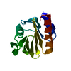
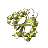
| Related structure data |  2molC 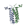 2mpnC  2moj C: citing same article ( |
|---|---|
| Similar structure data | |
| Other databases |


-Links
 PDBj
PDBj- Assembly
| Deposited unit | 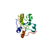
| |||||||||
|---|---|---|---|---|---|---|---|---|---|---|
| 1 |
| |||||||||
| NMR ensembles |
|
-Components
| #1: Protein | Mass: 11716.311 Da / Num. of mol.: 1 Source method: isolated from a genetically manipulated source Source: (gene. exp.) |
|---|
-Experimental details
-Experiment
| Experiment | Method: SOLUTION NMR | ||||||||||||||||||||||||||||||||||||
|---|---|---|---|---|---|---|---|---|---|---|---|---|---|---|---|---|---|---|---|---|---|---|---|---|---|---|---|---|---|---|---|---|---|---|---|---|---|
| NMR experiment |
|
 HSQC
HSQC- Sample preparation
| Details | Contents: 2 mM [U-100% 13C; U-100% 15N] YgaP, 90% H2O/10% D2O Solvent system: 90% H2O/10% D2O |
|---|---|
| Sample | Conc.: 2 mM / Component: entity-1 / Isotopic labeling: [U-100% 13C; U-100% 15N] |
| Sample conditions | Ionic strength: 0 / pH: 7 / Pressure: ambient / Temperature: 303 K |
-NMR measurement
| NMR spectrometer | Type: Bruker Avance / Manufacturer: Bruker / Model: AVANCE / Field strength: 700 MHz |
|---|
- Processing
| NMR software |
| ||||||||||||
|---|---|---|---|---|---|---|---|---|---|---|---|---|---|
| Refinement | Method: simulated annealing / Software ordinal: 1 | ||||||||||||
| NMR representative | Selection criteria: fewest violations | ||||||||||||
| NMR ensemble | Conformer selection criteria: target function / Conformers calculated total number: 100 / Conformers submitted total number: 10 |
