 Movie
Movie Controller

[English] 日本語
 Yorodumi
- PDB-2ml2: Solution Structure of AlgE6R2 subunit from the Azotobacter vinela... -
+ Open data

- Basic information
| Entry | Database: PDB / ID: 2ml2 | ||||||
|---|---|---|---|---|---|---|---|
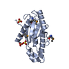
| Title | Solution Structure of AlgE6R2 subunit from the Azotobacter vinelandii Mannuronan C5-epimerase | ||||||
 Components Components | Poly(beta-D-mannuronate) C5 epimerase 6 | ||||||
 Keywords Keywords | ISOMERASE / Alginate C-5 epimerase / mannuronan C-5 epimerase / R-module | ||||||
| Function / homology |  Function and homology information Function and homology informationmannuronan 5-epimerase / alginic acid biosynthetic process / isomerase activity / calcium ion binding / : Similarity search - Function | ||||||
| Biological species |  Azotobacter vinelandii (bacteria) Azotobacter vinelandii (bacteria) | ||||||
| Method | SOLUTION NMR / simulated annealing | ||||||
| Model details | lowest energy, model1 | ||||||
 Authors Authors | Buchinger, E. / Wimmer, R. / Aachmann, F.L. | ||||||
 Citation Citation | Journal: J.Biol.Chem. / Year: 2014 Title: Structural and Functional Characterization of the R-modules in Alginate C-5 Epimerases AlgE4 and AlgE6 from Azotobacter vinelandii Authors: Buchinger, E. / Knudsen, D.H. / Behrens, M.A. / Pedersen, J.S. / Aarstad, O.A. / Tndervik, A. / Valla, S. / Skjak-Brk, G. / Wimmer, R. / Aachmann, F.L. #1: Journal: Biomol.Nmr Assign. / Year: 2011 Title: 1H, 13C and 15N resonances of the AlgE62 subunit from Azotobacter vinelandii mannuronan C5-epimerase Authors: Andreassen, T. / Buchinger, E. / Skjak-Brk, G. / Valla, S. / Aachmann, F.L. | ||||||
| History |
|
- Structure visualization
| Structure viewer | Molecule: MolmilJmol/JSmol |
|---|
- Downloads & links
-Download
| PDBx/mmCIF format | 2ml2.cif.gz | 893.8 KB | Display | PDBx/mmCIF format |
|---|---|---|---|---|
| PDB format | pdb2ml2.ent.gz | 746.4 KB | Display | PDB format |
| PDBx/mmJSON format | 2ml2.json.gz | Tree view | PDBx/mmJSON format | |
| Others |  Other downloads Other downloads |
-Validation report
| Arichive directory | https://data.pdbj.org/pub/pdb/validation_reports/ml/2ml2ftp://data.pdbj.org/pub/pdb/validation_reports/ml/2ml2 | HTTPS FTP |
|---|
-Related structure data
| Related structure data |  2ml1C  2ml3C C: citing same article ( |
|---|---|
| Similar structure data | |
| Other databases |
|


-Links
 PDBj
PDBj- Assembly
| Deposited unit | 
| |||||||||
|---|---|---|---|---|---|---|---|---|---|---|
| 1 |
| |||||||||
| NMR ensembles |
|
-Components
| #1: Protein | Mass: 16547.721 Da / Num. of mol.: 1 / Fragment: UNP residues 534-694 / Mutation: H72Q Source method: isolated from a genetically manipulated source Source: (gene. exp.) Azotobacter vinelandii (bacteria) / Strain: 354 Eubacteria / Gene: algE6 / Production host: References: UniProt: Q9ZFH0, Isomerases; Racemases and epimerases; Acting on carbohydrates and derivatives |
|---|---|
| #2: Chemical | ChemComp-CA /   Mass: 40.078 Da / Num. of mol.: 6 / Source method: obtained synthetically / Formula: Ca Mass: 40.078 Da / Num. of mol.: 6 / Source method: obtained synthetically / Formula: Ca |
-Experimental details
-Experiment
| Experiment | Method: SOLUTION NMR Details: Structure of the second R-module of AlgE6 from Azotobacter vinelandii | ||||||||||||||||||||||||||||||||||||||||||||||||||||||||||||||||||||||||
|---|---|---|---|---|---|---|---|---|---|---|---|---|---|---|---|---|---|---|---|---|---|---|---|---|---|---|---|---|---|---|---|---|---|---|---|---|---|---|---|---|---|---|---|---|---|---|---|---|---|---|---|---|---|---|---|---|---|---|---|---|---|---|---|---|---|---|---|---|---|---|---|---|---|
| NMR experiment |
|
 HSQC
HSQC- Sample preparation
| Details |
| ||||||||||||||||||||||||||||||||||||||||||||||||||||||||||||||||||||||||||||||||||||
|---|---|---|---|---|---|---|---|---|---|---|---|---|---|---|---|---|---|---|---|---|---|---|---|---|---|---|---|---|---|---|---|---|---|---|---|---|---|---|---|---|---|---|---|---|---|---|---|---|---|---|---|---|---|---|---|---|---|---|---|---|---|---|---|---|---|---|---|---|---|---|---|---|---|---|---|---|---|---|---|---|---|---|---|---|---|
| Sample |
| ||||||||||||||||||||||||||||||||||||||||||||||||||||||||||||||||||||||||||||||||||||
| Sample conditions | Ionic strength: 0.165 / pH: 6.9 / Pressure: 1 atm / Temperature: 298 K |
-NMR measurement
| NMR spectrometer | Type: Bruker Avance / Manufacturer: Bruker / Model: AVANCE / Field strength: 600 MHz |
|---|
- Processing
| NMR software |
| ||||||||||||||||||||||||||||
|---|---|---|---|---|---|---|---|---|---|---|---|---|---|---|---|---|---|---|---|---|---|---|---|---|---|---|---|---|---|
| Refinement | Method: simulated annealing / Software ordinal: 1 | ||||||||||||||||||||||||||||
| NMR constraints | NOE constraints total: 3579 / NOE intraresidue total count: 2316 / NOE long range total count: 397 / NOE medium range total count: 106 / NOE sequential total count: 760 / Protein chi angle constraints total count: 0 / Protein other angle constraints total count: 0 / Protein phi angle constraints total count: 52 / Protein psi angle constraints total count: 52 | ||||||||||||||||||||||||||||
| NMR representative | Selection criteria: lowest energy | ||||||||||||||||||||||||||||
| NMR ensemble | Conformer selection criteria: target function / Conformers calculated total number: 100 / Conformers submitted total number: 20 / Representative conformer: 1 |
