 Movie
Movie Controller

[English] 日本語
 Yorodumi
- PDB-2mak: Solution structure of the STIM1 CC1-CC2 homodimer in complex with... -
+ Open data

- Basic information
| Entry | Database: PDB / ID: 2mak | ||||||
|---|---|---|---|---|---|---|---|
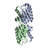
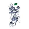
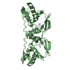
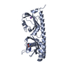
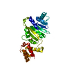
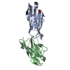
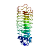
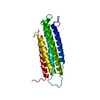
| Title | Solution structure of the STIM1 CC1-CC2 homodimer in complex with two Orai1 C-terminal domains. | ||||||
 Components Components |
| ||||||
 Keywords Keywords | SIGNALING PROTEIN/TRANSPORT PROTEIN / STIM1 / Orai1 / coiled-coil / Orai1 C-terminal domain / TRANSPORT PROTEIN / SIGNALING PROTEIN-TRANSPORT PROTEIN complex | ||||||
| Function / homology |  Function and homology information Function and homology informationstore-operated calcium entry / activation of store-operated calcium channel activity / positive regulation of adenylate cyclase activity / regulation of store-operated calcium entry / enamel mineralization / cortical endoplasmic reticulum / store-operated calcium channel activity / calcineurin-NFAT signaling cascade / calcium-ion regulated exocytosis / Elevation of cytosolic Ca2+ levels ...store-operated calcium entry / activation of store-operated calcium channel activity / positive regulation of adenylate cyclase activity / regulation of store-operated calcium entry / enamel mineralization / cortical endoplasmic reticulum / store-operated calcium channel activity / calcineurin-NFAT signaling cascade / calcium-ion regulated exocytosis / Elevation of cytosolic Ca2+ levels / microtubule plus-end binding / positive regulation of calcium ion transport / regulation of calcium ion transport / plasma membrane raft / detection of calcium ion / Ion homeostasis / sarcoplasmic reticulum membrane / calcium channel complex / Antigen activates B Cell Receptor (BCR) leading to generation of second messengers / cell periphery / calcium channel regulator activity / intracellular calcium ion homeostasis / calcium ion transmembrane transport / calcium channel activity / positive regulation of angiogenesis / protease binding / basolateral plasma membrane / microtubule / phospholipase C-activating G protein-coupled receptor signaling pathway / adaptive immune response / calmodulin binding / membrane raft / calcium ion binding / endoplasmic reticulum membrane / endoplasmic reticulum / membrane / identical protein binding / plasma membrane Similarity search - Function | ||||||
| Biological species |  Homo sapiens (human) Homo sapiens (human) | ||||||
| Method | SOLUTION NMR / simulated annealing | ||||||
| Model details | lowest energy, model1 | ||||||
 Authors Authors | Stathopulos, P.B. / Ikura, M. | ||||||
 Citation Citation | Journal: Nat Commun / Year: 2013 Title: STIM1/Orai1 coiled-coil interplay in the regulation of store-operated calcium entry. Authors: Stathopulos, P.B. / Schindl, R. / Fahrner, M. / Zheng, L. / Gasmi-Seabrook, G.M. / Muik, M. / Romanin, C. / Ikura, M. | ||||||
| History |
|
- Structure visualization
| Structure viewer | Molecule: MolmilJmol/JSmol |
|---|
- Downloads & links
-Download
| PDBx/mmCIF format | 2mak.cif.gz | 1.5 MB | Display | PDBx/mmCIF format |
|---|---|---|---|---|
| PDB format | pdb2mak.ent.gz | 1.3 MB | Display | PDB format |
| PDBx/mmJSON format | 2mak.json.gz | Tree view | PDBx/mmJSON format | |
| Others |  Other downloads Other downloads |
-Validation report
| Arichive directory | https://data.pdbj.org/pub/pdb/validation_reports/ma/2makftp://data.pdbj.org/pub/pdb/validation_reports/ma/2mak | HTTPS FTP |
|---|
-Related structure data
| Related structure data | 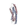 2majC C: citing same article ( |
|---|---|
| Similar structure data | |
| Other databases |
|


-Links
 PDBj
PDBj


- Assembly
| Deposited unit | 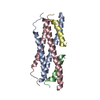
| |||||||||
|---|---|---|---|---|---|---|---|---|---|---|
| 1 |
| |||||||||
| NMR ensembles |
|
-Components
| #1: Protein | Mass: 9702.989 Da / Num. of mol.: 2 / Fragment: UNP residues 312-387 Source method: isolated from a genetically manipulated source Source: (gene. exp.) Homo sapiens (human) / Gene: STIM1, GOK / Production host:  #2: Protein/peptide | Mass: 2654.804 Da / Num. of mol.: 2 / Fragment: Orai1 C-terminal domain, UNP residues 272-292 Source method: isolated from a genetically manipulated source Source: (gene. exp.) Homo sapiens (human) / Gene: ORAI1, CRACM1, TMEM142A / Production host: |
|---|
-Experimental details
-Experiment
| Experiment | Method: SOLUTION NMR | ||||||||||||||||||||||||||||||||||||||||||||||||||||||||
|---|---|---|---|---|---|---|---|---|---|---|---|---|---|---|---|---|---|---|---|---|---|---|---|---|---|---|---|---|---|---|---|---|---|---|---|---|---|---|---|---|---|---|---|---|---|---|---|---|---|---|---|---|---|---|---|---|---|
| NMR experiment |
|
 HSQC
HSQC- Sample preparation
| Details |
| ||||||||||||||||||||||||||||||||||||||||||||||||||||||||||||||||||||||||||||||||||||
|---|---|---|---|---|---|---|---|---|---|---|---|---|---|---|---|---|---|---|---|---|---|---|---|---|---|---|---|---|---|---|---|---|---|---|---|---|---|---|---|---|---|---|---|---|---|---|---|---|---|---|---|---|---|---|---|---|---|---|---|---|---|---|---|---|---|---|---|---|---|---|---|---|---|---|---|---|---|---|---|---|---|---|---|---|---|
| Sample |
| ||||||||||||||||||||||||||||||||||||||||||||||||||||||||||||||||||||||||||||||||||||
| Sample conditions | Ionic strength: 0.02 / pH: 5.5 / Pressure: ambient / Temperature: 308 K |
-NMR measurement
| NMR spectrometer |
|
|---|
- Processing
| NMR software |
| ||||||||||||||||||||||||||||||||||||
|---|---|---|---|---|---|---|---|---|---|---|---|---|---|---|---|---|---|---|---|---|---|---|---|---|---|---|---|---|---|---|---|---|---|---|---|---|---|
| Refinement | Method: simulated annealing / Software ordinal: 1 Details: Used the RECOORD scripts in CNS to water refine the CYANA-determined structures (Nederveen et al., Proteins. 2005 Jun 1;59(4):662-72). | ||||||||||||||||||||||||||||||||||||
| NMR constraints | NOE constraints total: 2760 / NOE intraresidue total count: 676 / NOE long range total count: 233 / NOE medium range total count: 944 / NOE sequential total count: 907 / Hydrogen bond constraints total count: 280 / Protein phi angle constraints total count: 176 / Protein psi angle constraints total count: 176 | ||||||||||||||||||||||||||||||||||||
| NMR representative | Selection criteria: lowest energy | ||||||||||||||||||||||||||||||||||||
| NMR ensemble | Conformer selection criteria: structures with the lowest energy Conformers calculated total number: 200 / Conformers submitted total number: 20 / Maximum torsion angle constraint violation: 4.13 ° / Maximum upper distance constraint violation: -0.22 Å Torsion angle constraint violation method: RECOORD in CNS (Proteins. 2005 Jun 1;59(4):662-72) | ||||||||||||||||||||||||||||||||||||
| NMR ensemble rms | Distance rms dev: 0.023 Å / Distance rms dev error: 0.001 Å |
