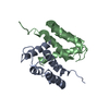
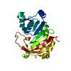
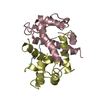
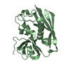
- PDB-2lux: Calcium saturated form of human C85M S100A1 mutant -
+
Open data
ID or keywords:
Loading...
-
Basic information
Entry
Database: PDB / ID: 2lux
Title
Calcium saturated form of human C85M S100A1 mutant
Components
Protein S100-A1
Keywords
METAL BINDING PROTEIN
Function / homology
Function and homology information
Regulation of TLR by endogenous ligand / MyD88 deficiency (TLR2/4) / S100 protein binding / M band / IRAK4 deficiency (TLR2/4) / MyD88:MAL(TIRAP) cascade initiated on plasma membrane / positive regulation of sprouting angiogenesis / regulation of heart contraction / substantia nigra development / sarcoplasmic reticulum ...Regulation of TLR by endogenous ligand / MyD88 deficiency (TLR2/4) / S100 protein binding / M band / IRAK4 deficiency (TLR2/4) / MyD88:MAL(TIRAP) cascade initiated on plasma membrane / positive regulation of sprouting angiogenesis / regulation of heart contraction / substantia nigra development / sarcoplasmic reticulum / vasodilation / Z disc / calcium-dependent protein binding / ATPase binding / ER-Phagosome pathway / intracellular signal transduction / calcium ion binding / negative regulation of transcription by RNA polymerase II / Golgi apparatus / protein homodimerization activity / protein-containing complex / mitochondrion / extracellular region / nucleoplasm / identical protein binding / nucleus / cytoplasm / cytosol Similarity search - Function
Protein S100-A1 / S-100/ICaBP type calcium binding protein signature. / S100/Calcium binding protein 7/8-like, conserved site / S100/CaBP-9k-type, calcium binding, subdomain / S-100/ICaBP type calcium binding domain / S-100/ICaBP type calcium binding domain / EF hand domain / EF-hand / Recoverin; domain 1 / EF-hand, calcium binding motif ...Protein S100-A1 / S-100/ICaBP type calcium binding protein signature. / S100/Calcium binding protein 7/8-like, conserved site / S100/CaBP-9k-type, calcium binding, subdomain / S-100/ICaBP type calcium binding domain / S-100/ICaBP type calcium binding domain / EF hand domain / EF-hand / Recoverin; domain 1 / EF-hand, calcium binding motif / EF-Hand 1, calcium-binding site / EF-hand calcium-binding domain. / EF-hand calcium-binding domain profile. / EF-hand domain / EF-hand domain pair / Orthogonal Bundle / Mainly Alpha Similarity search - Domain/homology
In the structure databanks used in Yorodumi, some data are registered as the other names, "COVID-19 virus" and "2019-nCoV". Here are the details of the virus and the list of structure data.
Jan 31, 2019. EMDB accession codes are about to change! (news from PDBe EMDB page)
EMDB accession codes are about to change! (news from PDBe EMDB page)
The allocation of 4 digits for EMDB accession codes will soon come to an end. Whilst these codes will remain in use, new EMDB accession codes will include an additional digit and will expand incrementally as the available range of codes is exhausted. The current 4-digit format prefixed with “EMD-” (i.e. EMD-XXXX) will advance to a 5-digit format (i.e. EMD-XXXXX), and so on. It is currently estimated that the 4-digit codes will be depleted around Spring 2019, at which point the 5-digit format will come into force.
The EM Navigator/Yorodumi systems omit the EMD- prefix.
Related info.:Q: What is EMD? / ID/Accession-code notation in Yorodumi/EM Navigator
Yorodumi is a browser for structure data from EMDB, PDB, SASBDB, etc.
This page is also the successor to EM Navigator detail page, and also detail information page/front-end page for Omokage search.
The word "yorodu" (or yorozu) is an old Japanese word meaning "ten thousand". "mi" (miru) is to see.
Related info.:EMDB / PDB / SASBDB / Comparison of 3 databanks / Yorodumi Search / Aug 31, 2016. New EM Navigator & Yorodumi / Yorodumi Papers / Jmol/JSmol / Function and homology information / Changes in new EM Navigator and Yorodumi
 Movie
Movie Controller


 Open data
 Basic information
 Components
 Keywords
 Function and homology information
 Homo sapiens (human)
 Authors
 Citation
 Structure visualization
 Downloads & links
 Other downloads
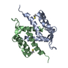


 PDBj
PDBj


 Assembly


 Mass: 40.078 Da / Num. of mol.: 4 / Source method: obtained synthetically / Formula: Ca
 HSQC
HSQC Sample preparation
 Processing