 Movie
Movie Controller

[English] 日本語
 Yorodumi
- PDB-2lr2: Designed IgG and lanthanide binding probe for solution NMR, MRI a... -
+ Open data

- Basic information
| Entry | Database: PDB / ID: 2lr2 | ||||||
|---|---|---|---|---|---|---|---|
| Title | Designed IgG and lanthanide binding probe for solution NMR, MRI and luminescence microscopy | ||||||
 Components Components | Immunoglobulin G-binding protein A | ||||||
 Keywords Keywords | DE NOVO PROTEIN / z domain / lanthanide binding tag | ||||||
| Function / homology | Immunoglobulin FC, subunit C / Single alpha-helices involved in coiled-coils or other helix-helix interfaces / Up-down Bundle / Mainly Alpha Function and homology information Function and homology information | ||||||
| Biological species | artificial gene (others) | ||||||
| Method | SOLUTION NMR / simulated annealing | ||||||
| Model details | lowest energy, model 1 | ||||||
 Authors Authors | Barb, A.W. / Ho, T.G. / Prestegard, J.H. | ||||||
 Citation Citation | Journal: Protein Sci. / Year: 2012 Title: Lanthanide binding and IgG affinity construct: Potential applications in solution NMR, MRI, and luminescence microscopy. Authors: Barb, A.W. / Ho, T.G. / Flanagan-Steet, H. / Prestegard, J.H. | ||||||
| History |
|
- Structure visualization
| Structure viewer | Molecule: MolmilJmol/JSmol |
|---|
- Downloads & links
-Download
| PDBx/mmCIF format | 2lr2.cif.gz | 55.2 KB | Display | PDBx/mmCIF format |
|---|---|---|---|---|
| PDB format | pdb2lr2.ent.gz | 41.3 KB | Display | PDB format |
| PDBx/mmJSON format | 2lr2.json.gz | Tree view | PDBx/mmJSON format | |
| Others |  Other downloads Other downloads |
-Validation report
| Summary document | 2lr2_validation.pdf.gz | 479.4 KB | Display | wwPDB validaton report |
|---|---|---|---|---|
| Full document | 2lr2_full_validation.pdf.gz | 511.7 KB | Display | |
| Data in XML | 2lr2_validation.xml.gz | 9.6 KB | Display | |
| Data in CIF | 2lr2_validation.cif.gz | 10.3 KB | Display | |
| Arichive directory | https://data.pdbj.org/pub/pdb/validation_reports/lr/2lr2ftp://data.pdbj.org/pub/pdb/validation_reports/lr/2lr2 | HTTPS FTP |
-Related structure data
| Similar structure data | |
|---|---|
| Other databases |


-Links
 PDBj
PDBj


- Assembly
| Deposited unit | 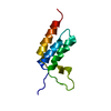
| |||||||||
|---|---|---|---|---|---|---|---|---|---|---|
| 1 |
| |||||||||
| NMR ensembles |
|
-Components
| #1: Antibody | Mass: 9875.544 Da / Num. of mol.: 1 Source method: isolated from a genetically manipulated source Source: (gene. exp.) artificial gene (others) / Production host:  |
|---|---|
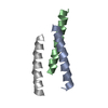
| Compound details | THE AUTHORS STATE THAT THE THREE HELICES ARE FROM THE Z DOMAIN WHICH IS A DERIVATIVE OF THE B ...THE AUTHORS STATE THAT THE THREE HELICES ARE FROM THE Z DOMAIN WHICH IS A DERIVATIVE |
-Experimental details
-Experiment
| Experiment | Method: SOLUTION NMR Details: This entry details the top 2 models from an xplor simulation to satisfy the orientation of a lanthanide-binding domain with the underlying Protein A-derived Z domain using paramagnetic NMR ...Details: This entry details the top 2 models from an xplor simulation to satisfy the orientation of a lanthanide-binding domain with the underlying Protein A-derived Z domain using paramagnetic NMR constraints. There is no explicit information regarding the placement of the domain-connecting loops, but these were included in the calculation and are presented to demonstrate the potential covalent restriction of the lanthanide-binding tag portion of the chimeric protein. | ||||||||||||||||||||
|---|---|---|---|---|---|---|---|---|---|---|---|---|---|---|---|---|---|---|---|---|---|
| NMR experiment |
|
 HSQC
HSQC- Sample preparation
| Details | Contents: 100-800 uM [U-98% 13C; U-98% 15N] Z-L2LBT, 90 % H2O, 10 % [U-2H] D2O, 90% H2O/10% D2O Solvent system: 90% H2O/10% D2O | ||||||||||||||||||||||||
|---|---|---|---|---|---|---|---|---|---|---|---|---|---|---|---|---|---|---|---|---|---|---|---|---|---|
| Sample |
| ||||||||||||||||||||||||
| Sample conditions | Ionic strength: 0.1 / pH: 8 / Pressure: ambient / Temperature: 273 K |
-NMR measurement
| NMR spectrometer |
|
|---|
- Processing
| NMR software |
| |||||||||
|---|---|---|---|---|---|---|---|---|---|---|
| Refinement | Method: simulated annealing / Software ordinal: 1 / Details: dihedral, PRE and PCS constraints used. | |||||||||
| NMR representative | Selection criteria: lowest energy | |||||||||
| NMR ensemble | Conformer selection criteria: structures with the lowest energy Conformers calculated total number: 50 / Conformers submitted total number: 2 |
