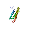
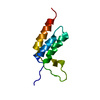
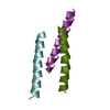
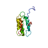
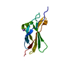
- PDB-4u7y: Structure of the complex of VPS4B MIT and IST1 MIM -
+
Open data
ID or keywords:
Loading...
-
Basic information
Entry
Database: PDB / ID: 4u7y
Title
Structure of the complex of VPS4B MIT and IST1 MIM
Components
IST1 homolog
Vacuolar protein sorting-associated protein 4B
Keywords
PROTEIN TRANSPORT / complex / MIM1
Function / homology
Function and homology information
protein depolymerization / late endosomal microautophagy / positive regulation of centriole elongation / late endosome to lysosome transport via multivesicular body sorting pathway / negative regulation of exosomal secretion / MIT domain binding / : / ESCRT III complex disassembly / cytoskeleton-dependent cytokinesis / nuclear membrane reassembly ...protein depolymerization / late endosomal microautophagy / positive regulation of centriole elongation / late endosome to lysosome transport via multivesicular body sorting pathway / negative regulation of exosomal secretion / MIT domain binding / : / ESCRT III complex disassembly / cytoskeleton-dependent cytokinesis / nuclear membrane reassembly / multivesicular body sorting pathway / vacuole organization / Sealing of the nuclear envelope (NE) by ESCRT-III / midbody abscission / regulation of centrosome duplication / positive regulation of exosomal secretion / membrane fission / multivesicular body assembly / ubiquitin-dependent protein catabolic process via the multivesicular body sorting pathway / establishment of blood-brain barrier / cholesterol transport / plasma membrane repair / endosome to lysosome transport via multivesicular body sorting pathway / Flemming body / regulation of mitotic spindle assembly / nucleus organization / vesicle-fusing ATPase / ATPase complex / endosomal transport / viral budding via host ESCRT complex / response to lipid / endoplasmic reticulum-Golgi intermediate compartment / mitotic metaphase chromosome alignment / positive regulation of proteolysis / canonical Wnt signaling pathway / nuclear pore / positive regulation of G2/M transition of mitotic cell cycle / autophagosome maturation / Endosomal Sorting Complex Required For Transport (ESCRT) / viral budding from plasma membrane / potassium ion transport / macroautophagy / establishment of protein localization / Budding and maturation of HIV virion / autophagy / spindle pole / azurophil granule lumen / intracellular protein localization / nuclear envelope / late endosome membrane / protein transport / angiogenesis / midbody / endosome / endosome membrane / cadherin binding / protein domain specific binding / cell division / centrosome / Neutrophil degranulation / chromatin / protein-containing complex binding / protein homodimerization activity / ATP hydrolysis activity / extracellular exosome / extracellular region / ATP binding / identical protein binding / nucleus / plasma membrane / cytosol / cytoplasm Similarity search - Function
Vacuolar protein sorting-associated protein 4, MIT domain / Phosphotransferase system, lactose/cellobiose-type IIA subunit / Vacuolar protein sorting-associated protein Ist1 / Vacuolar protein sorting-associated protein IST1-like / Regulator of Vps4 activity in the MVB pathway / MIT (microtubule interacting and transport) domain / MIT domain superfamily / Vps4 oligomerisation, C-terminal / MIT domain / Microtubule Interacting and Trafficking molecule domain ...Vacuolar protein sorting-associated protein 4, MIT domain / Phosphotransferase system, lactose/cellobiose-type IIA subunit / Vacuolar protein sorting-associated protein Ist1 / Vacuolar protein sorting-associated protein IST1-like / Regulator of Vps4 activity in the MVB pathway / MIT (microtubule interacting and transport) domain / MIT domain superfamily / Vps4 oligomerisation, C-terminal / MIT domain / Microtubule Interacting and Trafficking molecule domain / : / Vps4 C terminal oligomerisation domain / AAA ATPase, AAA+ lid domain / AAA+ lid domain / ATPase, AAA-type, conserved site / AAA-protein family signature. / Methane Monooxygenase Hydroxylase; Chain G, domain 1 / ATPase family associated with various cellular activities (AAA) / ATPase, AAA-type, core / ATPases associated with a variety of cellular activities / AAA+ ATPase domain / Up-down Bundle / P-loop containing nucleoside triphosphate hydrolase / Mainly Alpha Similarity search - Domain/homology
National Institutes of Health/National Institute of General Medical Sciences (NIH/NIGMS)
GM095769
United States
Citation
Journal: J.Biol.Chem. / Year: 2015 Title: Distinct Mechanisms of Recognizing Endosomal Sorting Complex Required for Transport III (ESCRT-III) Protein IST1 by Different Microtubule Interacting and Trafficking (MIT) Domains. Authors: Guo, E.Z. / Xu, Z.
In the structure databanks used in Yorodumi, some data are registered as the other names, "COVID-19 virus" and "2019-nCoV". Here are the details of the virus and the list of structure data.
Jan 31, 2019. EMDB accession codes are about to change! (news from PDBe EMDB page)
EMDB accession codes are about to change! (news from PDBe EMDB page)
The allocation of 4 digits for EMDB accession codes will soon come to an end. Whilst these codes will remain in use, new EMDB accession codes will include an additional digit and will expand incrementally as the available range of codes is exhausted. The current 4-digit format prefixed with “EMD-” (i.e. EMD-XXXX) will advance to a 5-digit format (i.e. EMD-XXXXX), and so on. It is currently estimated that the 4-digit codes will be depleted around Spring 2019, at which point the 5-digit format will come into force.
The EM Navigator/Yorodumi systems omit the EMD- prefix.
Related info.:Q: What is EMD? / ID/Accession-code notation in Yorodumi/EM Navigator
Yorodumi is a browser for structure data from EMDB, PDB, SASBDB, etc.
This page is also the successor to EM Navigator detail page, and also detail information page/front-end page for Omokage search.
The word "yorodu" (or yorozu) is an old Japanese word meaning "ten thousand". "mi" (miru) is to see.
Related info.:EMDB / PDB / SASBDB / Comparison of 3 databanks / Yorodumi Search / Aug 31, 2016. New EM Navigator & Yorodumi / Yorodumi Papers / Jmol/JSmol / Function and homology information / Changes in new EM Navigator and Yorodumi
 Movie
Movie Controller


 Open data
 Basic information
 Components
 Keywords
 Function and homology information
 Homo sapiens (human)
 X-RAY DIFFRACTION /
  Authors
 United States, 1items
  Citation
 Structure visualization
 Downloads & links
 Other downloads


 PDBj
PDBj


 Assembly

 Mass: 18.015 Da / Num. of mol.: 6 / Source method: isolated from a natural source / Formula: H2O
 Sample preparation
 Processing