[English] 日本語
Yorodumi
- PDB-2lfg: Solution structure of the human prolactin receptor ecd domain d2 -

+
Open data


ID or keywords:

Loading...

-
Basic information

Entry
Database: PDB / ID: 2lfg
TitleSolution structure of the human prolactin receptor ecd domain d2
ComponentsProlactin receptor
KeywordsHORMONE RECEPTOR / Extracellular domain / Cytokine receptor
Function / homology
Function and homology information


prolactin receptor activity / regulation of epithelial cell differentiation / prostate gland growth / mammary gland epithelial cell differentiation / steroid biosynthetic process / activation of Janus kinase activity / cellular response to granulocyte macrophage colony-stimulating factor stimulus / activation of transmembrane receptor protein tyrosine kinase activity / cytokine binding / Prolactin receptor signaling ...prolactin receptor activity / regulation of epithelial cell differentiation / prostate gland growth / mammary gland epithelial cell differentiation / steroid biosynthetic process / activation of Janus kinase activity / cellular response to granulocyte macrophage colony-stimulating factor stimulus / activation of transmembrane receptor protein tyrosine kinase activity / cytokine binding / Prolactin receptor signaling / peptide hormone binding / cell surface receptor signaling pathway via JAK-STAT / mammary gland alveolus development / regulation of cell adhesion / positive regulation of B cell proliferation / Growth hormone receptor signaling / embryo implantation / positive regulation of protein autophosphorylation / lactation / endosome lumen / response to bacterium / cytokine-mediated signaling pathway / positive regulation of cold-induced thermogenesis / receptor complex / external side of plasma membrane / lipid binding / positive regulation of cell population proliferation / negative regulation of apoptotic process / protein kinase binding / cell surface / extracellular region / metal ion binding / plasma membrane
Similarity search - Function
Long hematopoietin receptor, single chain, conserved site / Long hematopoietin receptor, single chain family signature. / Growth hormone/erythropoietin receptor, ligand binding / Erythropoietin receptor, ligand binding / Fibronectin type 3 domain / Fibronectin type-III domain profile. / Fibronectin type III / Fibronectin type III superfamily / Immunoglobulins / Immunoglobulin-like fold ...Long hematopoietin receptor, single chain, conserved site / Long hematopoietin receptor, single chain family signature. / Growth hormone/erythropoietin receptor, ligand binding / Erythropoietin receptor, ligand binding / Fibronectin type 3 domain / Fibronectin type-III domain profile. / Fibronectin type III / Fibronectin type III superfamily / Immunoglobulins / Immunoglobulin-like fold / Immunoglobulin-like / Sandwich / Mainly Beta
Similarity search - Domain/homology
Biological speciesHomo sapiens (human)
MethodSOLUTION NMR / simulated annealing
Model detailslowest energy, model 1
AuthorsDagil, R. / Knudsen, M.J. / Kragelund, B.B. / O'Shea, C. / Teilum, K.
CitationJournal: Structure / Year: 2012
Title: The WSXWS Motif in Cytokine Receptors Is a Molecular Switch Involved in Receptor Activation: Insight from Structures of the Prolactin Receptor
Authors: Dagil, R. / Knudsen, M.J. / Olsen, J.G. / O'Shea, C. / Franzmann, M. / Goffin, V. / Teilum, K. / Breinholt, J. / Kragelund, B.B.
History
DepositionJun 30, 2011Deposition site: BMRB / Processing site: RCSB
Revision 1.0Feb 15, 2012Provider: repository / Type: Initial release
Revision 1.1Feb 22, 2012Group: Database references
Revision 1.2Jun 14, 2023Group: Data collection / Database references / Other
Category: database_2 / pdbx_database_status ...database_2 / pdbx_database_status / pdbx_nmr_software / pdbx_nmr_spectrometer / struct_ref_seq_dif
Item: _database_2.pdbx_DOI / _database_2.pdbx_database_accession ..._database_2.pdbx_DOI / _database_2.pdbx_database_accession / _pdbx_database_status.status_code_nmr_data / _pdbx_nmr_software.name / _pdbx_nmr_spectrometer.model / _struct_ref_seq_dif.details

-
Structure visualization

Structure viewerMolecule:
MolmilJmol/JSmol

Downloads & links

-
Assembly

Deposited unit
A: Prolactin receptor


Theoretical massNumber of molelcules
Total (without water)13,3431
Polymers13,3431
Non-polymers00
Water0
1


  • Idetical with deposited unit
  • defined by author
TypeNameSymmetry operationNumber
identity operation1_555x,y,z1
NMR ensembles
DataCriteria
Number of conformers (submitted / calculated)20 / 100
RepresentativeModel #1lowest energy

-
Components

#1: Protein Prolactin receptor / / PRL-R


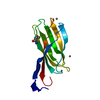
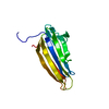
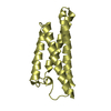
Mass: 13343.341 Da / Num. of mol.: 1 / Fragment: Fibronectin type-III 2 domain residues 99-210
Source method: isolated from a genetically manipulated source
Source: (gene. exp.) Homo sapiens (human) / Gene: PRLR / Production host: Escherichia coli (E. coli) / Strain (production host): BL21(DE3) / References: UniProt: P16471

-
Experimental details

-
Experiment

ExperimentMethod: SOLUTION NMR
NMR experiment
Conditions-IDExperiment-IDSolution-IDType
1112D 1H-15N HSQC
1212D 1H-13C HSQC
1313D C(CO)NH
1413D HNCO
1513D HNCA
1613D HN(CA)CB
1713D HN(CO)CA
1813D H(CCO)NH
1913D CBCA(CO)NH
11013D HN(CA)CO
11113D 1H-15N NOESY
11213D 1H-15N TOCSY
11313D (H)CCH-TOCSY
11413D 1H-13C NOESY aliphatic
11513D 1H-13C NOESY aromatic
11622D 1H-13C HSQC aromatic

-
Sample preparation

Details
Solution-IDContentsSolvent system
10.5 mM [U-100% 13C; U-100% 15N] protein, 10 mM sodium phosphate, 10 mM TCEP, 1 mM DSS, 0.02 % sodium azide, 90% H2O/10% D2O90% H2O/10% D2O
20.5 mM [U-100% 13C(1)] protein, 10 mM sodium phosphate, 10 mM TCEP, 0.02 % sodium azide, 1 mM DSS, 100% D2O100% D2O
Sample
Conc. (mg/ml)ComponentIsotopic labelingSolution-ID
0.5 mMprotein-1[U-100% 13C; U-100% 15N]1
10 mMsodium phosphate-21
10 mMTCEP-31
1 mMDSS-41
0.02 %sodium azide-51
0.5 mMprotein-6[U-100% 13C(1)]2
10 mMsodium phosphate-72
10 mMTCEP-82
0.02 %sodium azide-92
1 mMDSS-102
Sample conditionspH: 7.4 / Pressure: ambient / Temperature: 298 K

-
NMR measurement

NMR spectrometer
TypeManufacturerModelField strength (MHz)Spectrometer-ID
Varian INOVAVarianINOVA8001
Bruker AvanceBrukerAVANCE8002

-
Processing

NMR software
NameVersionDeveloperClassification
AutoAssign2.3.0Zimmerman, Moseley, Kulikowski and Montelionechemical shift assignment
CYANA2.2.1Guntert, Mumenthaler and Wuthrichchemical shift assignment
X-PLOR NIH2.25Schwieters, Kuszewski, Tjandra and Clorestructure solution
TALOSCornilescu, Delaglio and Baxgeometry optimization
NMRDrawDelaglio, Grzesiek, Vuister, Zhu, Pfeifer and Baxprocessing
CCPN2.1.5CCPNpeak picking
ARIA2.3.2Linge, O'Donoghue and Nilgeswater refinement
X-PLOR NIHSchwieters, Kuszewski, Tjandra and Clorerefinement
RefinementMethod: simulated annealing / Software ordinal: 1
NMR constraintsNOE constraints total: 1562 / NOE intraresidue total count: 345 / NOE long range total count: 583 / NOE medium range total count: 634 / Protein phi angle constraints total count: 54 / Protein psi angle constraints total count: 55
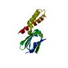
NMR representativeSelection criteria: lowest energy
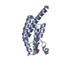
NMR ensembleConformers calculated total number: 100 / Conformers submitted total number: 20 / Maximum lower distance constraint violation: 0.25 Å / Maximum upper distance constraint violation: 0.34 Å

+
About Yorodumi

-
News

-
Feb 9, 2022. New format data for meta-information of EMDB entries

New format data for meta-information of EMDB entries

  • Version 3 of the EMDB header file is now the official format.
  • The previous official version 1.9 will be removed from the archive.

Related info.:EMDB header

External links:wwPDB to switch to version 3 of the EMDB data model

-
Aug 12, 2020. Covid-19 info

Covid-19 info

URL: https://pdbj.org/emnavi/covid19.php

New page: Covid-19 featured information page in EM Navigator.

Related info.:Covid-19 info / Mar 5, 2020. Novel coronavirus structure data

+
Mar 5, 2020. Novel coronavirus structure data

Novel coronavirus structure data

Related info.:Yorodumi Speices / Aug 12, 2020. Covid-19 info

External links:COVID-19 featured content - PDBj / Molecule of the Month (242):Coronavirus Proteases

+
Jan 31, 2019. EMDB accession codes are about to change! (news from PDBe EMDB page)

EMDB accession codes are about to change! (news from PDBe EMDB page)

  • The allocation of 4 digits for EMDB accession codes will soon come to an end. Whilst these codes will remain in use, new EMDB accession codes will include an additional digit and will expand incrementally as the available range of codes is exhausted. The current 4-digit format prefixed with “EMD-” (i.e. EMD-XXXX) will advance to a 5-digit format (i.e. EMD-XXXXX), and so on. It is currently estimated that the 4-digit codes will be depleted around Spring 2019, at which point the 5-digit format will come into force.
  • The EM Navigator/Yorodumi systems omit the EMD- prefix.

Related info.:Q: What is EMD? / ID/Accession-code notation in Yorodumi/EM Navigator

External links:EMDB Accession Codes are Changing Soon! / Contact to PDBj

+
Jul 12, 2017. Major update of PDB

Major update of PDB

  • wwPDB released updated PDB data conforming to the new PDBx/mmCIF dictionary.
  • This is a major update changing the version number from 4 to 5, and with Remediation, in which all the entries are updated.
  • In this update, many items about electron microscopy experimental information are reorganized (e.g. em_software).
  • Now, EM Navigator and Yorodumi are based on the updated data.

External links:wwPDB Remediation / Enriched Model Files Conforming to OneDep Data Standards Now Available in the PDB FTP Archive

-
Yorodumi

Thousand views of thousand structures

  • Yorodumi is a browser for structure data from EMDB, PDB, SASBDB, etc.
  • This page is also the successor to EM Navigator detail page, and also detail information page/front-end page for Omokage search.
  • The word "yorodu" (or yorozu) is an old Japanese word meaning "ten thousand". "mi" (miru) is to see.

Related info.:EMDB / PDB / SASBDB / Comparison of 3 databanks / Yorodumi Search / Aug 31, 2016. New EM Navigator & Yorodumi / Yorodumi Papers / Jmol/JSmol / Function and homology information / Changes in new EM Navigator and Yorodumi

Read more