 ムービー
ムービー コントローラー


+ データを開く

- 基本情報
基本情報
| 登録情報 | データベース: PDB / ID: 2l5h | ||||||
|---|---|---|---|---|---|---|---|
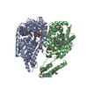
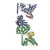
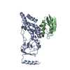
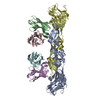
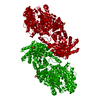
| タイトル | Solution Structure of the H189Q mutant of the Enzyme I dimer Using Residual Dipolar Couplings and Small Angle X-Ray Scattering | ||||||
 要素 要素 | Phosphoenolpyruvate-protein phosphotransferase | ||||||
 キーワード キーワード | TRANSFERASE / protein / dimer | ||||||
| 機能・相同性 |  機能・相同性情報 機能・相同性情報phosphoenolpyruvate-protein phosphotransferase / phosphoenolpyruvate-protein phosphotransferase activity / N-acetylglucosamine transport / phosphoenolpyruvate-dependent sugar phosphotransferase system / kinase activity / metal ion binding / identical protein binding / cytosol 類似検索 - 分子機能 | ||||||
| 生物種 |  | ||||||
| 手法 | 溶液NMR /  溶液散乱 / simulated annealing 溶液散乱 / simulated annealing | ||||||
| Model details | lowest energy, model 1 | ||||||
 データ登録者 データ登録者 | Takayama, Y.D. / Schwieters, C.D. / Grishaev, A. / Guirlando, R. / Clore, G. | ||||||
 引用 引用 | ジャーナル: J.Am.Chem.Soc. / 年: 2011 タイトル: Combined Use of Residual Dipolar Couplings and Solution X-ray Scattering To Rapidly Probe Rigid-Body Conformational Transitions in a Non-phosphorylatable Active-Site Mutant of the 128 kDa Enzyme I Dimer. 著者: Takayama, Y. / Schwieters, C.D. / Grishaev, A. / Ghirlando, R. / Clore, G.M. | ||||||
| 履歴 |
|
- 構造の表示
構造の表示
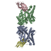
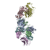
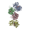
| 構造ビューア | 分子: MolmilJmol/JSmol |
|---|
- ダウンロードとリンク
-ダウンロード
| PDBx/mmCIF形式 | 2l5h.cif.gz | 759.1 KB | 表示 | PDBx/mmCIF形式 |
|---|---|---|---|---|
| PDB形式 | pdb2l5h.ent.gz | 639.8 KB | 表示 | PDB形式 |
| PDBx/mmJSON形式 | 2l5h.json.gz | ツリー表示 | PDBx/mmJSON形式 | |
| その他 |  その他のダウンロード その他のダウンロード |
-検証レポート
| 文書・要旨 | 2l5h_validation.pdf.gz | 386.9 KB | 表示 | wwPDB検証レポート |
|---|---|---|---|---|
| 文書・詳細版 | 2l5h_full_validation.pdf.gz | 526.5 KB | 表示 | |
| XML形式データ | 2l5h_validation.xml.gz | 66.2 KB | 表示 | |
| CIF形式データ | 2l5h_validation.cif.gz | 93.2 KB | 表示 | |
| アーカイブディレクトリ | https://data.pdbj.org/pub/pdb/validation_reports/l5/2l5hftp://data.pdbj.org/pub/pdb/validation_reports/l5/2l5h | HTTPS FTP |
-関連構造データ
| 関連構造データ | |
|---|---|
| 類似構造データ |


-リンク
 PDBj
PDBj
- 集合体
集合体
| 登録構造単位 | 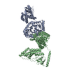
| |||||||||
|---|---|---|---|---|---|---|---|---|---|---|
| 1 |
| |||||||||
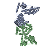
| NMR アンサンブル |
|
-要素
| #1: タンパク質 | 分子量: 63409.328 Da / 分子数: 2 / 由来タイプ: 組換発現 / 由来: (組換発現) 参照: UniProt: P08839, phosphoenolpyruvate-protein phosphotransferase |
|---|
-実験情報
-実験
| 実験 |
| |||
|---|---|---|---|---|
| NMR実験 | タイプ: TROSY-based 1H-15N correlation spectroscopy |
- 試料調製
試料調製
| 詳細 | 内容: 20 mM TRIS-1, 100 mM sodium chloride-2, 10 mM DTT-3, 4 mM MgCl2-4, 1 mM EDTA-5, 10 % D2O-6, 0.15 mM EI dimer-8, 90% H2O/10% D2O 溶媒系: 90% H2O/10% D2O | ||||||||||||||||||||||||
|---|---|---|---|---|---|---|---|---|---|---|---|---|---|---|---|---|---|---|---|---|---|---|---|---|---|
| 試料 |
| ||||||||||||||||||||||||
| 試料状態 | pH: 7.4 / 圧: ambient / 温度: 310 K |
-データ収集
| NMRスペクトロメーター | タイプ: Bruker DRX / 製造業者: Bruker / モデル: DRX / 磁場強度: 800 MHz |
|---|---|
| Soln scatter | タイプ: x-ray Buffer name: 20 mM Tris, 100 mM NaCl, 10 mM DTT, 4 mM MgCl2, 1 mM EDTA, 1 tablet protease inhibitor cocktail (SigmaFAST S8830) Conc. range: 5 mg/ml Data reduction software list: Mar_Detector, Igor, Primus, Gnom Detector specific: ECT division, Argonne National Laboratory 検出器タイプ: Gold CCD / Mean guiner radius: 4.1 nm / Mean guiner radius esd: 0.1 nm / Num. of time frames: 20 / Protein length: 15.5 / Sample pH: 7.4 / Source beamline: 12-IDC / Source class: Y / Source type: APS / 温度: 298 K |
- 解析
解析
| NMR software |
| ||||||||||||
|---|---|---|---|---|---|---|---|---|---|---|---|---|---|
| 精密化 | 手法: simulated annealing / ソフトェア番号: 1 | ||||||||||||
| NMR constraints | Protein chi angle constraints total count: 72 / Protein other angle constraints total count: 46 / Protein phi angle constraints total count: 32 / Protein psi angle constraints total count: 30 | ||||||||||||
| 代表構造 | 選択基準: lowest energy | ||||||||||||
| NMRアンサンブル | コンフォーマー選択の基準: target function / 計算したコンフォーマーの数: 120 / 登録したコンフォーマーの数: 2 | ||||||||||||
| Soln scatter model | 手法: simulated annealing / コンフォーマー選択の基準: lowest energy 詳細: The initial structure of the H189Q mutant of the EI dimer was taken from the calculated structures of the wildtype EI dimer summarized in PDB entry 2KX9. Throughout the structure ...詳細: The initial structure of the H189Q mutant of the EI dimer was taken from the calculated structures of the wildtype EI dimer summarized in PDB entry 2KX9. Throughout the structure determination, the backbone atomic coordinates of the c-terminus of each EI subunit (residues 262-573) were held fixed in space, while the two subdomains of each n-terminus (residues 25-142 in the alpha subdomain, and residues 1-21 and 147-254 in the alpha-beta subdomain) were treated as rigid bodies. Coordinates in the linker regions (residues 22-24, 143-146, and 255-261) and interfactial sidechains were allowed varying degrees of freedom during the calculation through the use of the internal variable module (IVM) of Xplor-NIH. Model 1 corresponds to the regularized mean of the 100 structures for which data was reported in the primary publication, with the B-factor column representing the per-atom spread (in B-factor units). Model 2 is the lowest energy structure. The calculated structural statistics for the original 96 structures and for the two reported structures are: MODEL 1: SAXS CHI2 Q->0.44: 0.63 SAXS CHI2 FULL RANGE: 0.80 RDC R-FACTOR: 18.76 RDC DA: 11.16 RDC RH: 0.59 MODEL 2: SAXS CHI2 Q->0.44: 0.50 SAXS CHI2 FULL RANGE: 0.72 RDC R-FACTOR: 19.18 RDC DA: 11.15 RDC RH: 0.60 AVERAGE OVER THE FULL 99-MEMBER ENSEMBLE: SAXS CHI2 Q->0.44: 0.58 +/- 0.10 SAXS CHI2 FULL RANGE: 0.76 +/- 0.16 RDC R-FACTOR: 19.02 +/- 0.37 RDC DA: 11.13+/- 0.19 RDC RH: 0.59 +/- 0.02 Entry fitting list: free wildtype EI structures, PDB ID 2KX9 Num. of conformers calculated: 120 / Num. of conformers submitted: 2 / 代表コンフォーマー: 1 Software author list: C.D. Schwieters, J.J. Kuszewski, N. Tjandra,G.M. Clore Software list: XPLOR-NIH |
