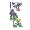
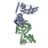
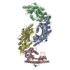
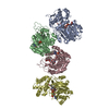
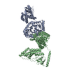
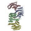
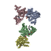
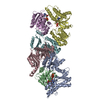
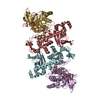
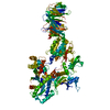
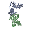
Journal: J Am Chem Soc / Year: 2010 Title: Solution structure of the 128 kDa enzyme I dimer from Escherichia coli and its 146 kDa complex with HPr using residual dipolar couplings and small- and wide-angle X-ray scattering. Authors: Charles D Schwieters / Jeong-Yong Suh / Alexander Grishaev / Rodolfo Ghirlando / Yuki Takayama / G Marius Clore / Abstract: The solution structures of free Enzyme I (EI, ∼128 kDa, 575 × 2 residues), the first enzyme in the bacterial phosphotransferase system, and its complex with HPr (∼146 kDa) have been solved using ...The solution structures of free Enzyme I (EI, ∼128 kDa, 575 × 2 residues), the first enzyme in the bacterial phosphotransferase system, and its complex with HPr (∼146 kDa) have been solved using novel methodology that makes use of prior structural knowledge (namely, the structures of the dimeric EIC domain and the isolated EIN domain both free and complexed to HPr), combined with residual dipolar coupling (RDC), small- (SAXS) and wide- (WAXS) angle X-ray scattering and small-angle neutron scattering (SANS) data. The calculational strategy employs conjoined rigid body/torsion/Cartesian simulated annealing, and incorporates improvements in calculating and refining against SAXS/WAXS data that take into account complex molecular shapes in the description of the solvent layer resulting in a better representation of the SAXS/WAXS data. The RDC data orient the symmetrically related EIN domains relative to the C(2) symmetry axis of the EIC dimer, while translational, shape, and size information is provided by SAXS/WAXS. The resulting structures are independently validated by SANS. Comparison of the structures of the free EI and the EI-HPr complex with that of the crystal structure of a trapped phosphorylated EI intermediate reveals large (∼70-90°) hinge body rotations of the two subdomains comprising the EIN domain, as well as of the EIN domain relative to the dimeric EIC domain. These large-scale interdomain motions shed light on the structural transitions that accompany the catalytic cycle of EI.
Name: 20mM TRIS buffer, 100 mM NaCl, 10 mM DTT, 4 mM MgCl2, 1 mM EDTA pH: 7.4 Comment: 1 tablet of protease inhibitor cocktail (SigmaFAST S8830) was added
Instrument name: Advanced Photon Source (APS) 12ID-C / City: Argonne, IL / 国: USA / Type of source: X-ray synchrotron / Wavelength: 0.06199 Å / Dist. spec. to detc.: 4 mm
Detector
Name: Gold CCD
Scan
Title: Phosphoenolpyruvate-protein phosphotransferase / Measurement date: Aug 23, 2010 / Cell temperature: 25 °C / Exposure time: 0.25 sec. / Number of frames: 20 / Unit: 1/A /
Min
Max
Q
0.0144
0.2208
Distance distribution function P(R)
Sofotware P(R): GNOM 5.0 / Number of points: 164 /
Min
Max
Q
0.014426
0.1953
P(R) point
1
164
R
0
148.6
Result
Type of curve: single_conc /
Experimental
Porod
MW
128 kDa
118 kDa
Volume
-
189 nm3
Guinier
P(R)
P(R) error
Guinier error
Forward scattering, I0
0.0290498
-
-
-
Radius of gyration, Rg
4.1 nm
4.2 nm
0.02
0.06
Max
Error
D
14.75
0.5
+
About Yorodumi
-
News
-
Feb 9, 2022. New format data for meta-information of EMDB entries
New format data for meta-information of EMDB entries
Version 3 of the EMDB header file is now the official format.
The previous official version 1.9 will be removed from the archive.
In the structure databanks used in Yorodumi, some data are registered as the other names, "COVID-19 virus" and "2019-nCoV". Here are the details of the virus and the list of structure data.
Jan 31, 2019. EMDB accession codes are about to change! (news from PDBe EMDB page)
EMDB accession codes are about to change! (news from PDBe EMDB page)
The allocation of 4 digits for EMDB accession codes will soon come to an end. Whilst these codes will remain in use, new EMDB accession codes will include an additional digit and will expand incrementally as the available range of codes is exhausted. The current 4-digit format prefixed with “EMD-” (i.e. EMD-XXXX) will advance to a 5-digit format (i.e. EMD-XXXXX), and so on. It is currently estimated that the 4-digit codes will be depleted around Spring 2019, at which point the 5-digit format will come into force.
The EM Navigator/Yorodumi systems omit the EMD- prefix.
Related info.:Q: What is EMD? / ID/Accession-code notation in Yorodumi/EM Navigator
Yorodumi is a browser for structure data from EMDB, PDB, SASBDB, etc.
This page is also the successor to EM Navigator detail page, and also detail information page/front-end page for Omokage search.
The word "yorodu" (or yorozu) is an old Japanese word meaning "ten thousand". "mi" (miru) is to see.
Related info.:EMDB / PDB / SASBDB / Comparison of 3 databanks / Yorodumi Search / Aug 31, 2016. New EM Navigator & Yorodumi / Yorodumi Papers / Jmol/JSmol / Function and homology information / Changes in new EM Navigator and Yorodumi
 Movie
Movie Controller

 Open data
 Basic information
 Sample
 Function and homology information
 Citation
 Structure visualization
 Downloads & links
 SASDCN3


 Search similar-shape structures of this assembly by Omokage search (details)


