 Movie
Movie Controller

[English] 日本語
 Yorodumi
- PDB-2l5h: Solution Structure of the H189Q mutant of the Enzyme I dimer Usin... -
+ Open data

- Basic information
| Entry | Database: PDB / ID: 2l5h | ||||||
|---|---|---|---|---|---|---|---|
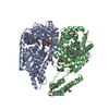
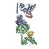
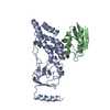
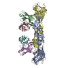
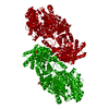
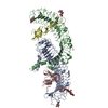
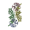
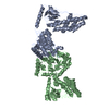
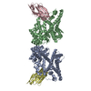
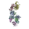
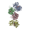
| Title | Solution Structure of the H189Q mutant of the Enzyme I dimer Using Residual Dipolar Couplings and Small Angle X-Ray Scattering | ||||||
 Components Components | Phosphoenolpyruvate-protein phosphotransferase | ||||||
 Keywords Keywords | TRANSFERASE / protein / dimer | ||||||
| Function / homology |  Function and homology information Function and homology informationphosphoenolpyruvate-protein phosphotransferase / phosphoenolpyruvate-protein phosphotransferase activity / N-acetylglucosamine transport / phosphoenolpyruvate-dependent sugar phosphotransferase system / kinase activity / metal ion binding / identical protein binding / cytosol Similarity search - Function | ||||||
| Biological species |  | ||||||
| Method | SOLUTION NMR /  SOLUTION SCATTERING / simulated annealing SOLUTION SCATTERING / simulated annealing | ||||||
| Model details | lowest energy, model 1 | ||||||
 Authors Authors | Takayama, Y.D. / Schwieters, C.D. / Grishaev, A. / Guirlando, R. / Clore, G. | ||||||
 Citation Citation | Journal: J.Am.Chem.Soc. / Year: 2011 Title: Combined Use of Residual Dipolar Couplings and Solution X-ray Scattering To Rapidly Probe Rigid-Body Conformational Transitions in a Non-phosphorylatable Active-Site Mutant of the 128 kDa Enzyme I Dimer. Authors: Takayama, Y. / Schwieters, C.D. / Grishaev, A. / Ghirlando, R. / Clore, G.M. | ||||||
| History |
|
- Structure visualization
| Structure viewer | Molecule: MolmilJmol/JSmol |
|---|
- Downloads & links
-Download
| PDBx/mmCIF format | 2l5h.cif.gz | 759.1 KB | Display | PDBx/mmCIF format |
|---|---|---|---|---|
| PDB format | pdb2l5h.ent.gz | 639.8 KB | Display | PDB format |
| PDBx/mmJSON format | 2l5h.json.gz | Tree view | PDBx/mmJSON format | |
| Others |  Other downloads Other downloads |
-Validation report
| Arichive directory | https://data.pdbj.org/pub/pdb/validation_reports/l5/2l5hftp://data.pdbj.org/pub/pdb/validation_reports/l5/2l5h | HTTPS FTP |
|---|
-Related structure data
| Related structure data | |
|---|---|
| Similar structure data |


-Links
 PDBj
PDBj
- Assembly
| Deposited unit | 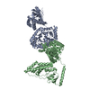
| |||||||||
|---|---|---|---|---|---|---|---|---|---|---|
| 1 |
| |||||||||
| NMR ensembles |
|
-Components
| #1: Protein | Mass: 63409.328 Da / Num. of mol.: 2 Source method: isolated from a genetically manipulated source Source: (gene. exp.) References: UniProt: P08839, phosphoenolpyruvate-protein phosphotransferase |
|---|
-Experimental details
-Experiment
| Experiment |
| |||
|---|---|---|---|---|
| NMR experiment | Type: TROSY-based 1H-15N correlation spectroscopy |
- Sample preparation
| Details | Contents: 20 mM TRIS-1, 100 mM sodium chloride-2, 10 mM DTT-3, 4 mM MgCl2-4, 1 mM EDTA-5, 10 % D2O-6, 0.15 mM EI dimer-8, 90% H2O/10% D2O Solvent system: 90% H2O/10% D2O | ||||||||||||||||||||||||
|---|---|---|---|---|---|---|---|---|---|---|---|---|---|---|---|---|---|---|---|---|---|---|---|---|---|
| Sample |
| ||||||||||||||||||||||||
| Sample conditions | pH: 7.4 / Pressure: ambient / Temperature: 310 K |
-Data collection
| NMR spectrometer | Type: Bruker DRX / Manufacturer: Bruker / Model: DRX / Field strength: 800 MHz |
|---|---|
| Soln scatter | Type: x-ray Buffer name: 20 mM Tris, 100 mM NaCl, 10 mM DTT, 4 mM MgCl2, 1 mM EDTA, 1 tablet protease inhibitor cocktail (SigmaFAST S8830) Conc. range: 5 mg/ml Data reduction software list: Mar_Detector, Igor, Primus, Gnom Detector specific: ECT division, Argonne National Laboratory Detector type: Gold CCD / Mean guiner radius: 4.1 nm / Mean guiner radius esd: 0.1 nm / Num. of time frames: 20 / Protein length: 15.5 / Sample pH: 7.4 / Source beamline: 12-IDC / Source class: Y / Source type: APS / Temperature: 298 K |
- Processing
| NMR software |
| ||||||||||||
|---|---|---|---|---|---|---|---|---|---|---|---|---|---|
| Refinement | Method: simulated annealing / Software ordinal: 1 | ||||||||||||
| NMR constraints | Protein chi angle constraints total count: 72 / Protein other angle constraints total count: 46 / Protein phi angle constraints total count: 32 / Protein psi angle constraints total count: 30 | ||||||||||||
| NMR representative | Selection criteria: lowest energy | ||||||||||||
| NMR ensemble | Conformer selection criteria: target function / Conformers calculated total number: 120 / Conformers submitted total number: 2 | ||||||||||||
| Soln scatter model | Method: simulated annealing / Conformer selection criteria: lowest energy Details: The initial structure of the H189Q mutant of the EI dimer was taken from the calculated structures of the wildtype EI dimer summarized in PDB entry 2KX9. Throughout the structure ...Details: The initial structure of the H189Q mutant of the EI dimer was taken from the calculated structures of the wildtype EI dimer summarized in PDB entry 2KX9. Throughout the structure determination, the backbone atomic coordinates of the c-terminus of each EI subunit (residues 262-573) were held fixed in space, while the two subdomains of each n-terminus (residues 25-142 in the alpha subdomain, and residues 1-21 and 147-254 in the alpha-beta subdomain) were treated as rigid bodies. Coordinates in the linker regions (residues 22-24, 143-146, and 255-261) and interfactial sidechains were allowed varying degrees of freedom during the calculation through the use of the internal variable module (IVM) of Xplor-NIH. Model 1 corresponds to the regularized mean of the 100 structures for which data was reported in the primary publication, with the B-factor column representing the per-atom spread (in B-factor units). Model 2 is the lowest energy structure. The calculated structural statistics for the original 96 structures and for the two reported structures are: MODEL 1: SAXS CHI2 Q->0.44: 0.63 SAXS CHI2 FULL RANGE: 0.80 RDC R-FACTOR: 18.76 RDC DA: 11.16 RDC RH: 0.59 MODEL 2: SAXS CHI2 Q->0.44: 0.50 SAXS CHI2 FULL RANGE: 0.72 RDC R-FACTOR: 19.18 RDC DA: 11.15 RDC RH: 0.60 AVERAGE OVER THE FULL 99-MEMBER ENSEMBLE: SAXS CHI2 Q->0.44: 0.58 +/- 0.10 SAXS CHI2 FULL RANGE: 0.76 +/- 0.16 RDC R-FACTOR: 19.02 +/- 0.37 RDC DA: 11.13+/- 0.19 RDC RH: 0.59 +/- 0.02 Entry fitting list: free wildtype EI structures, PDB ID 2KX9 Num. of conformers calculated: 120 / Num. of conformers submitted: 2 / Representative conformer: 1 Software author list: C.D. Schwieters, J.J. Kuszewski, N. Tjandra,G.M. Clore Software list: XPLOR-NIH |
