 Movie
Movie Controller


+ Open data

- Basic information
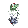
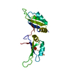
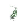
| Entry | Database: PDB / ID: 2l48 | ||||||
|---|---|---|---|---|---|---|---|
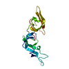
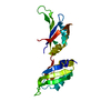
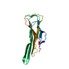
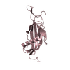
| Title | Solution structure of the PlyG cell wall binding domain | ||||||
 Components Components | N-acetylmuramoyl-L-alanine amidase | ||||||
 Keywords Keywords | Carbohydrate-Binding Protein / Bacillus anthracis gamma- phage endolysin / PlyG / cell wall binding domain / homodimer / ACT-type domain | ||||||
| Function / homology |  Function and homology information Function and homology informationN-acetylmuramoyl-L-alanine amidase / establishment of competence for transformation / N-acetylmuramoyl-L-alanine amidase activity / peptidoglycan turnover / sporulation resulting in formation of a cellular spore / viral release from host cell by cytolysis / peptidoglycan catabolic process / cell wall organization / defense response to bacterium Similarity search - Function | ||||||
| Biological species |  Bacillus phage Gamma (virus) Bacillus phage Gamma (virus) | ||||||
| Method | SOLUTION NMR / AUTOMATED METHODS WERE USED FOR BACKBONE CHEMICAL SHIFT ASSIGNMENT, ITERATIVE NOE REFINEMENT. FINAL STRUCTURES WERE OBTAINED BY MOLECULAR DYNAMICS IN EXPLICIT SOLVENT, AUTOMATED METHODS WERE USED FOR BACKBONE CHEMICAL SHIFT ASSIGNMENT, ITERATIVE NOE REFINEMENT. FINAL STRUCTURES WERE OBTAINED BY MOLECULAR DYNAMICS IN EXPLICIT SOLVENT | ||||||
 Authors Authors | Volkman, B.F. / Dias, J.S. / Peterson, F.C. | ||||||
 Citation Citation | Journal: To be Published Title: Tbd Authors: Dias, J.S. / Peterson, F.C. / Volkman, B.F. | ||||||
| History |
|
- Structure visualization
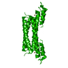
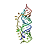
| Structure viewer | Molecule: MolmilJmol/JSmol |
|---|
- Downloads & links
-Download
| PDBx/mmCIF format | 2l48.cif.gz | 1 MB | Display | PDBx/mmCIF format |
|---|---|---|---|---|
| PDB format | pdb2l48.ent.gz | 911.2 KB | Display | PDB format |
| PDBx/mmJSON format | 2l48.json.gz | Tree view | PDBx/mmJSON format | |
| Others |  Other downloads Other downloads |
-Validation report
| Arichive directory | https://data.pdbj.org/pub/pdb/validation_reports/l4/2l48ftp://data.pdbj.org/pub/pdb/validation_reports/l4/2l48 | HTTPS FTP |
|---|
-Related structure data
| Related structure data | |
|---|---|
| Similar structure data |


-Links
 PDBj
PDBj- Assembly
| Deposited unit | 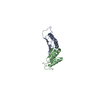
| |||||||||
|---|---|---|---|---|---|---|---|---|---|---|
| 1 |
| |||||||||
| NMR ensembles |
|
-Components
| #1: Protein | Mass: 9447.730 Da / Num. of mol.: 2 / Fragment: cell wall binding domain, residues 151-233 Source method: isolated from a genetically manipulated source Source: (gene. exp.) Bacillus phage Gamma (virus) / Gene: GAMMALSU_0017, GAMMAUSAM_0017, PlyG / Plasmid: pQE30 / Production host:  |
|---|
-Experimental details
-Experiment
| Experiment | Method: SOLUTION NMR | ||||||||||||||||
|---|---|---|---|---|---|---|---|---|---|---|---|---|---|---|---|---|---|
| NMR experiment |
|
- Sample preparation
| Details | Contents: 1.25 mM [U-100% 13C; U-100% 15N] PlyG, 20 mM [U-99% 2H] Bis-Tris, 90% H2O, 10% D2O Solvent system: 90% H2O/10% D2O | ||||||||||||
|---|---|---|---|---|---|---|---|---|---|---|---|---|---|
| Sample |
| ||||||||||||
| Sample conditions | Ionic strength: 13 / pH: 6 / Pressure: AMBIENT / Temperature: 298 K |
-NMR measurement
| NMR spectrometer | Type: Bruker DRX / Manufacturer: Bruker / Model: DRX / Field strength: 600 MHz |
|---|
- Processing
| NMR software |
| ||||||||||||||||||||||||||||
|---|---|---|---|---|---|---|---|---|---|---|---|---|---|---|---|---|---|---|---|---|---|---|---|---|---|---|---|---|---|
| Refinement | Method: AUTOMATED METHODS WERE USED FOR BACKBONE CHEMICAL SHIFT ASSIGNMENT, ITERATIVE NOE REFINEMENT. FINAL STRUCTURES WERE OBTAINED BY MOLECULAR DYNAMICS IN EXPLICIT SOLVENT, AUTOMATED METHODS WERE ...Method: AUTOMATED METHODS WERE USED FOR BACKBONE CHEMICAL SHIFT ASSIGNMENT, ITERATIVE NOE REFINEMENT. FINAL STRUCTURES WERE OBTAINED BY MOLECULAR DYNAMICS IN EXPLICIT SOLVENT, AUTOMATED METHODS WERE USED FOR BACKBONE CHEMICAL SHIFT ASSIGNMENT, ITERATIVE NOE REFINEMENT. FINAL STRUCTURES WERE OBTAINED BY MOLECULAR DYNAMICS IN EXPLICIT SOLVENT Software ordinal: 1 Details: HOMODIMER STRUCTURES ARE BASED ON A TOTAL OF 3966 NOE CONSTRAINTS (704 INTRA, 726 SEQUENTIAL, 958 MEDIUM and 1442 INTRAMONOMER LONG RANGE AND 136 INTERMONOMER CONSTRAINTS) AND 220 PHI AND ...Details: HOMODIMER STRUCTURES ARE BASED ON A TOTAL OF 3966 NOE CONSTRAINTS (704 INTRA, 726 SEQUENTIAL, 958 MEDIUM and 1442 INTRAMONOMER LONG RANGE AND 136 INTERMONOMER CONSTRAINTS) AND 220 PHI AND PSI DIHEDRAL ANGLE CONSTRAINTS. CONSTRAINTS WERE ASSIGNED AND VALIDATED IN ONE MONOMER AND THEN DUPLICATD TO GENERATE A SYMMETRY RELATED CONSTRAINT IN THE SECOND MONOMER. CONSTRAINT TOTALS LISTED ABOVE INCLUDE CONTRAINTS FROM BOTH MONOMERS, HOMODIMER STRUCTURES ARE BASED ON A TOTAL OF 3966 NOE CONSTRAINTS (704 INTRA, 726 SEQUENTIAL, 958 MEDIUM and 1422 INTRAMONOMER LONG RANGE AND 136 INTERMONOMER CONSTRAINTS) AND 220 PHI AND PSI DIHEDRAL ANGLE CONSTRAINTS. CONSTRAINTS WERE ASSIGNED AND VALIDATED IN ONE MONOMER AND THEN DUPLICATD TO GENERATE A SYMMETRY RELATED CONSTRAINT IN THE SECOND MONOMER. CONSTRAINT TOTALS LISTED ABOVE INCLUDE CONTRAINTS FROM BOTH MONOMERS | ||||||||||||||||||||||||||||
| NMR constraints | NOE constraints total: 3966 / NOE intraresidue total count: 704 / NOE long range total count: 1578 / NOE medium range total count: 958 / NOE sequential total count: 726 | ||||||||||||||||||||||||||||
| NMR representative | Selection criteria: lowest energy | ||||||||||||||||||||||||||||
| NMR ensemble | Conformer selection criteria: target function / Conformers calculated total number: 100 / Conformers submitted total number: 20 |
 Xplor-NIH