 Movie
Movie Controller

[English] 日本語
 Yorodumi
- PDB-2hh7: Crystal Structure of Cu(I) bound CsoR from Mycobacterium tuberculosis. -
+ Open data

- Basic information
| Entry | Database: PDB / ID: 2hh7 | ||||||
|---|---|---|---|---|---|---|---|
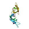
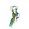
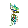
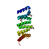
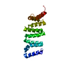
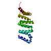
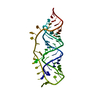
| Title | Crystal Structure of Cu(I) bound CsoR from Mycobacterium tuberculosis. | ||||||
 Components Components | Hypothetical protein CsoR | ||||||
 Keywords Keywords | UNKNOWN FUNCTION / 4-helix bundle | ||||||
| Function / homology |  Function and homology information Function and homology informationcopper ion sensor activity / response to silver ion / DNA-binding transcription repressor activity / response to copper ion / protein-DNA complex / transcription cis-regulatory region binding / copper ion binding / negative regulation of DNA-templated transcription / DNA binding / plasma membrane / cytoplasm Similarity search - Function | ||||||
| Biological species |   Mycobacterium tuberculosis (bacteria) Mycobacterium tuberculosis (bacteria) | ||||||
| Method |  X-RAY DIFFRACTION / SYNCHROTRON / MAD / Resolution: 2.55 Å X-RAY DIFFRACTION / SYNCHROTRON / MAD / Resolution: 2.55 Å | ||||||
 Authors Authors | Sacchettini, J.C. / Ramesh, A. | ||||||
 Citation Citation | Journal: Nat.Chem.Biol. / Year: 2007 Title: CsoR is a novel Mycobacterium tuberculosis copper-sensing transcriptional regulator. Authors: Liu, T. / Ramesh, A. / Ma, Z. / Ward, S.K. / Zhang, L. / George, G.N. / Talaat, A.M. / Sacchettini, J.C. / Giedroc, D.P. | ||||||
| History |
|
- Structure visualization
| Structure viewer | Molecule: MolmilJmol/JSmol |
|---|
- Downloads & links
-Download
| PDBx/mmCIF format | 2hh7.cif.gz | 29.5 KB | Display | PDBx/mmCIF format |
|---|---|---|---|---|
| PDB format | pdb2hh7.ent.gz | 19.3 KB | Display | PDB format |
| PDBx/mmJSON format | 2hh7.json.gz | Tree view | PDBx/mmJSON format | |
| Others |  Other downloads Other downloads |
-Validation report
| Arichive directory | https://data.pdbj.org/pub/pdb/validation_reports/hh/2hh7ftp://data.pdbj.org/pub/pdb/validation_reports/hh/2hh7 | HTTPS FTP |
|---|
-Related structure data
| Similar structure data |
|---|

-Links
 PDBj
PDBj- Assembly
| Deposited unit | 
| ||||||||
|---|---|---|---|---|---|---|---|---|---|
| 1 | 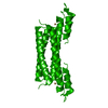
| ||||||||
| 2 | 
| ||||||||
| Unit cell |
| ||||||||
| Details | The second part of the biological assembly is generated by the symmetry operation : x, x-y-1, -z-1/3 |
-Components
| #1: Protein | Mass: 12817.682 Da / Num. of mol.: 1 Source method: isolated from a genetically manipulated source Source: (gene. exp.) Mycobacterium tuberculosis (bacteria) / Production host: |
|---|---|
| #2: Chemical | ChemComp-CU1 /   Mass: 63.546 Da / Num. of mol.: 1 / Source method: obtained synthetically / Formula: Cu Mass: 63.546 Da / Num. of mol.: 1 / Source method: obtained synthetically / Formula: Cu |
| #3: Water | ChemComp-HOH /  Mass: 18.015 Da / Num. of mol.: 20 / Source method: isolated from a natural source / Formula: H2O Mass: 18.015 Da / Num. of mol.: 20 / Source method: isolated from a natural source / Formula: H2O |
-Experimental details
-Experiment
| Experiment | Method: X-RAY DIFFRACTION / Number of used crystals: 1 |
|---|
- Sample preparation
| Crystal | Density Matthews: 2.18 Å3/Da / Density % sol: 43.66 % |
|---|---|
| Crystal grow | Temperature: 290 K / Method: vapor diffusion, hanging drop / pH: 5.6 Details: 30% Peg4000,0.1Msodium citrate tribasic dihydrate, 0.2M ammonium acetate, pH 5.6, VAPOR DIFFUSION, HANGING DROP, temperature 290K |
-Data collection
| Diffraction |
| |||||||||||||||
|---|---|---|---|---|---|---|---|---|---|---|---|---|---|---|---|---|
| Diffraction source |
| |||||||||||||||
| Detector |
| |||||||||||||||
| Radiation | Protocol: MAD / Monochromatic (M) / Laue (L): M / Scattering type: x-ray | |||||||||||||||
| Radiation wavelength |
| |||||||||||||||
| Reflection | Resolution: 2.55→50 Å / Num. all: 4011 / Num. obs: 3987 / % possible obs: 99.4 % / Observed criterion σ(F): 2 / Observed criterion σ(I): 4.24 | |||||||||||||||
| Reflection shell | Highest resolution: 2.55 Å / % possible all: 99.2 |

- Processing
| Software |
| ||||||||||||||||||||
|---|---|---|---|---|---|---|---|---|---|---|---|---|---|---|---|---|---|---|---|---|---|
| Refinement | Method to determine structure: MAD / Resolution: 2.55→20 Å / σ(F): 4.24 / Stereochemistry target values: Engh & Huber
| ||||||||||||||||||||
| Refinement step | Cycle: LAST / Resolution: 2.55→20 Å
| ||||||||||||||||||||
| Refine LS restraints |
| ||||||||||||||||||||
| LS refinement shell | Highest resolution: 2.55 Å
|
