 Movie
Movie Controller


+ Open data

- Basic information
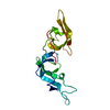
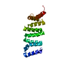
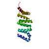
| Entry | Database: PDB / ID: 1qqr | ||||||
|---|---|---|---|---|---|---|---|
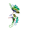
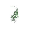
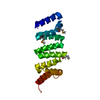
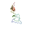
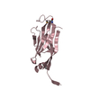
| Title | CRYSTAL STRUCTURE OF STREPTOKINASE DOMAIN B | ||||||
 Components Components | STREPTOKINASE DOMAIN B | ||||||
 Keywords Keywords | HYDROLASE ACTIVATOR / NON-PROTEOLYTIC / PLASMINOGEN ACTIVATION / FIBRINOLYSIS | ||||||
| Function / homology |  Function and homology information Function and homology information | ||||||
| Biological species |  Streptococcus dysgalactiae subsp. equisimilis (bacteria) Streptococcus dysgalactiae subsp. equisimilis (bacteria) | ||||||
| Method |  X-RAY DIFFRACTION / SYNCHROTRON / MOLECULAR REPLACEMENT / Resolution: 2.3 Å X-RAY DIFFRACTION / SYNCHROTRON / MOLECULAR REPLACEMENT / Resolution: 2.3 Å | ||||||
 Authors Authors | Spraggon, G. / Zhang, X.X. / Ponting, C.P. / Fox, V.F. / Phillips, C. / Smith, R.A.G. / Jones, E.Y. / Dobson, C. / Stuart, D.I. | ||||||
 Citation Citation | Journal: To be Published Title: Crystal Structure of Streptokinse Domain B Authors: Spraggon, G. / Zhang, X.X. / Ponting, C.P. / Fox, V.F. / Phillips, C. / Smith, R.A.G. / Jones, E.Y. / Dobson, C. / Stuart, D.I. | ||||||
| History |
|
- Structure visualization
| Structure viewer | Molecule: MolmilJmol/JSmol |
|---|
- Downloads & links
-Download
| PDBx/mmCIF format | 1qqr.cif.gz | 127.2 KB | Display | PDBx/mmCIF format |
|---|---|---|---|---|
| PDB format | pdb1qqr.ent.gz | 101.1 KB | Display | PDB format |
| PDBx/mmJSON format | 1qqr.json.gz | Tree view | PDBx/mmJSON format | |
| Others |  Other downloads Other downloads |
-Validation report
| Arichive directory | https://data.pdbj.org/pub/pdb/validation_reports/qq/1qqrftp://data.pdbj.org/pub/pdb/validation_reports/qq/1qqr | HTTPS FTP |
|---|
-Related structure data
| Related structure data |  1bmlS S: Starting model for refinement |
|---|---|
| Similar structure data |

-Links
 PDBj
PDBj- Assembly
| Deposited unit | 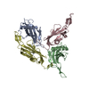
| ||||||||||
|---|---|---|---|---|---|---|---|---|---|---|---|
| 1 | 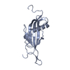
| ||||||||||
| 2 | 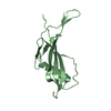
| ||||||||||
| 3 | 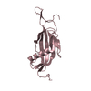
| ||||||||||
| 4 | 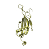
| ||||||||||
| Unit cell |
|
-Components
| #1: Protein | Mass: 16021.934 Da / Num. of mol.: 4 / Fragment: B-DOMAIN / Source method: isolated from a natural source Source: (natural) Streptococcus dysgalactiae subsp. equisimilis (bacteria)Species: Streptococcus dysgalactiae / Strain: subsp. equisimilis / References: UniProt: P00779 #2: Water | ChemComp-HOH / |  Mass: 18.015 Da / Num. of mol.: 337 / Source method: isolated from a natural source / Formula: H2O Mass: 18.015 Da / Num. of mol.: 337 / Source method: isolated from a natural source / Formula: H2O |
|---|
-Experimental details
-Experiment
| Experiment | Method: X-RAY DIFFRACTION / Number of used crystals: 1 |
|---|
- Sample preparation
| Crystal | Density Matthews: 2.9 Å3/Da / Density % sol: 57.63 % |
|---|
-Data collection
| Diffraction | Mean temperature: 150 K |
|---|---|
| Diffraction source | Source: SYNCHROTRON / Site: EMBL/DESY, HAMBURG  / Beamline: X11 / Wavelength: 1 / Beamline: X11 / Wavelength: 1 |
| Detector | Type: MARRESEARCH / Detector: IMAGE PLATE |
| Radiation | Protocol: SINGLE WAVELENGTH / Monochromatic (M) / Laue (L): M / Scattering type: x-ray |
| Radiation wavelength | Wavelength: 1 Å / Relative weight: 1 |
| Reflection | Resolution: 2.3→50 Å / Num. all: 34029 / Num. obs: 31237 / Observed criterion σ(F): 0 / Observed criterion σ(I): 0 / Biso Wilson estimate: 44.7 Å2 / Rmerge(I) obs: 0.078 |
- Processing
| Software |
| ||||||||||||||||||||||||||||||||||||||||||||||||||||||||||||
|---|---|---|---|---|---|---|---|---|---|---|---|---|---|---|---|---|---|---|---|---|---|---|---|---|---|---|---|---|---|---|---|---|---|---|---|---|---|---|---|---|---|---|---|---|---|---|---|---|---|---|---|---|---|---|---|---|---|---|---|---|---|
| Refinement | Method to determine structure: MOLECULAR REPLACEMENT Starting model: 1BML Resolution: 2.3→50 Å / Rfactor Rfree error: 0.006 / Isotropic thermal model: RESTRAINED / Cross valid method: THROUGHOUT / σ(F): 0 / σ(I): 0 / Stereochemistry target values: ENGH & HUBER / Details: MAXIMUM LIKELIHOOD REFINEMENT
| ||||||||||||||||||||||||||||||||||||||||||||||||||||||||||||
| Solvent computation | Solvent model: FLAT MODEL / Bsol: 68.098 Å2 / ksol: 0.35326 e/Å3 | ||||||||||||||||||||||||||||||||||||||||||||||||||||||||||||
| Displacement parameters | Biso mean: 47.9 Å2
| ||||||||||||||||||||||||||||||||||||||||||||||||||||||||||||
| Refine analyze |
| ||||||||||||||||||||||||||||||||||||||||||||||||||||||||||||
| Refinement step | Cycle: LAST / Resolution: 2.3→50 Å
| ||||||||||||||||||||||||||||||||||||||||||||||||||||||||||||
| Refine LS restraints |
| ||||||||||||||||||||||||||||||||||||||||||||||||||||||||||||
| LS refinement shell | Resolution: 2.3→2.44 Å / Rfactor Rfree error: 0.025 / Total num. of bins used: 6
| ||||||||||||||||||||||||||||||||||||||||||||||||||||||||||||
| Xplor file | Serial no: 1 / Param file: PROTEIN_REP.PARAM / Topol file: PROTEIN.TOP |
