 Movie
Movie Controller


+ Open data

- Basic information
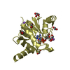
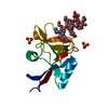
| Entry | Database: PDB / ID: 2l47 | ||||||
|---|---|---|---|---|---|---|---|
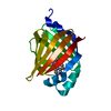
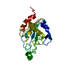
| Title | Solution structure of the PlyG catalytic domain | ||||||
 Components Components | N-acetylmuramoyl-L-alanine amidase | ||||||
 Keywords Keywords | HYDROLASE / Bacillus anthracis gamma-phage endolysin / PlyG / amidase-2 family / Zn-dependent peptidoglycan amidase / catalytic domain | ||||||
| Function / homology |  Function and homology information Function and homology informationN-acetylmuramoyl-L-alanine amidase / establishment of competence for transformation / N-acetylmuramoyl-L-alanine amidase activity / peptidoglycan turnover / sporulation resulting in formation of a cellular spore / viral release from host cell by cytolysis / peptidoglycan catabolic process / cell wall organization / defense response to bacterium Similarity search - Function | ||||||
| Biological species |  Bacillus phage Gamma (virus) Bacillus phage Gamma (virus) | ||||||
| Method | SOLUTION NMR / AUTOMATED METHODS WERE USED FOR BACKBONE CHEMICAL SHIFT ASSIGNMENT, ITERATIVE NOE REFINEMENT. FINAL STRUCTURES WERE OBTAINED BY MOLECULAR DYNAMICS IN EXPLICIT SOLVENT | ||||||
 Authors Authors | Volkman, B.F. / Dias, J.S. / Peterson, F.C. | ||||||
 Citation Citation | Journal: To be Published Title: Tbd Authors: Dias, J.S. / Peterson, F.C. / Volkman, B.F. | ||||||
| History |
|
- Structure visualization
| Structure viewer | Molecule: MolmilJmol/JSmol |
|---|
- Downloads & links
-Download
| PDBx/mmCIF format | 2l47.cif.gz | 1.1 MB | Display | PDBx/mmCIF format |
|---|---|---|---|---|
| PDB format | pdb2l47.ent.gz | 930.9 KB | Display | PDB format |
| PDBx/mmJSON format | 2l47.json.gz | Tree view | PDBx/mmJSON format | |
| Others |  Other downloads Other downloads |
-Validation report
| Arichive directory | https://data.pdbj.org/pub/pdb/validation_reports/l4/2l47ftp://data.pdbj.org/pub/pdb/validation_reports/l4/2l47 | HTTPS FTP |
|---|
-Related structure data
| Related structure data | |
|---|---|
| Similar structure data |


-Links
 PDBj
PDBj- Assembly
| Deposited unit | 
| |||||||||
|---|---|---|---|---|---|---|---|---|---|---|
| 1 |
| |||||||||
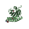
| NMR ensembles |
|
-Components
| #1: Protein | Mass: 18554.877 Da / Num. of mol.: 1 / Fragment: catalytic domain, residues 1-165 Source method: isolated from a genetically manipulated source Source: (gene. exp.) Bacillus phage Gamma (virus) / Gene: PlyG, GAMMALSU_0017, GAMMAUSAM_0017 / Plasmid: pBAD18 / Production host:  References: UniProt: Q8LTE6, N-acetylmuramoyl-L-alanine amidase |
|---|---|
| #2: Chemical | ChemComp-ZN /   Mass: 65.409 Da / Num. of mol.: 1 / Source method: obtained synthetically / Formula: Zn Mass: 65.409 Da / Num. of mol.: 1 / Source method: obtained synthetically / Formula: Zn |
-Experimental details
-Experiment
| Experiment | Method: SOLUTION NMR | ||||||||||||||||||||
|---|---|---|---|---|---|---|---|---|---|---|---|---|---|---|---|---|---|---|---|---|---|
| NMR experiment |
|
- Sample preparation
| Details | Contents: 1 mM [U-100% 13C; U-100% 15N] PlyG, 10 mM [U-99% 2H] Bis-Tris, 90% H2O, 10% D2O Solvent system: 90% H2O/10% D2O | ||||||||||||
|---|---|---|---|---|---|---|---|---|---|---|---|---|---|
| Sample |
| ||||||||||||
| Sample conditions | Ionic strength: 13 / pH: 6 / Pressure: AMBIENT / Temperature: 303 K |
-NMR measurement
| NMR spectrometer | Type: Bruker DRX / Manufacturer: Bruker / Model: DRX / Field strength: 600 MHz |
|---|
- Processing
| NMR software |
| ||||||||||||||||||||||||||||
|---|---|---|---|---|---|---|---|---|---|---|---|---|---|---|---|---|---|---|---|---|---|---|---|---|---|---|---|---|---|
| Refinement | Method: AUTOMATED METHODS WERE USED FOR BACKBONE CHEMICAL SHIFT ASSIGNMENT, ITERATIVE NOE REFINEMENT. FINAL STRUCTURES WERE OBTAINED BY MOLECULAR DYNAMICS IN EXPLICIT SOLVENT Software ordinal: 1 Details: STRUCTURES ARE BASED ON A TOTAL OF 3503 NOE CONSTRAINTS (634 INTRA, 594 SEQUENTIAL, 750 MEDIUM and 1525 LONG RANGE CONSTRAINTS) AND 216 PHI AND PSI DIHEDRAL ANGLE CONSTRAINTS. | ||||||||||||||||||||||||||||
| NMR constraints | NOE constraints total: 3503 / NOE intraresidue total count: 634 / NOE long range total count: 1525 / NOE medium range total count: 750 / NOE sequential total count: 594 | ||||||||||||||||||||||||||||
| NMR representative | Selection criteria: lowest energy | ||||||||||||||||||||||||||||
| NMR ensemble | Conformer selection criteria: target function / Conformers calculated total number: 100 / Conformers submitted total number: 20 |
 Xplor-NIH