 Movie
Movie Controller

[English] 日本語
 Yorodumi
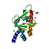
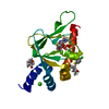
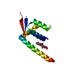
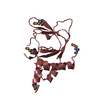
- PDB-2kke: Solution NMR Structure of a dimeric protein of unknown function f... -
+ Open data

- Basic information
| Entry | Database: PDB / ID: 2kke | ||||||
|---|---|---|---|---|---|---|---|
| Title | Solution NMR Structure of a dimeric protein of unknown function from Methanobacterium thermoautotrophicum, Northeast Structural Genomics Consortium Target TR5 | ||||||
 Components Components | Uncharacterized protein | ||||||
 Keywords Keywords | Structural Genomics / Unknown function / Protein NMR / NORTHEAST STRUCTURAL GENOMICS CONSORTIUM (NESG) / Target TR5 / PSI-2 / Protein Structure Initiative / Methanothermobacter thermautotrophicus / O26567_METTH / Northeast Structural Genomics Consortium / NESG | ||||||
| Function / homology | Arc Repressor Mutant - #60 / Arc Repressor Mutant / Orthogonal Bundle / Mainly Alpha / CopG family transcriptional regulator Function and homology information Function and homology information | ||||||
| Biological species |   Methanothermobacter thermautotrophicus str. Delta H (archaea) Methanothermobacter thermautotrophicus str. Delta H (archaea) | ||||||
| Method | SOLUTION NMR / simulated annealing | ||||||
| Model details | lowest energy, model 1 | ||||||
 Authors Authors | Swapna, G.V.T. / Gunsalus, X. / Huang, L. / Xiao, K. / Everett, J.K. / Acton, T.B. / Montelione, G.T. / Northeast Structural Genomics Consortium (NESG) | ||||||
 Citation Citation | Journal: To be Published Title: NMR Solution Structure of a putative uncharacterized protein from Methanobacterium thermoautotrophicum, Northeast Structural Genomics Consortium Target:TR5 Authors: Swapna, G.V.T. / Gunsalus, X. / Huang, L. / Xiao, K. / Everett, J.K. / Acton, T.B. / Montelione, G.T. | ||||||
| History |
|
- Structure visualization
| Structure viewer | Molecule: MolmilJmol/JSmol |
|---|
- Downloads & links
-Download
| PDBx/mmCIF format | 2kke.cif.gz | 730.3 KB | Display | PDBx/mmCIF format |
|---|---|---|---|---|
| PDB format | pdb2kke.ent.gz | 619.7 KB | Display | PDB format |
| PDBx/mmJSON format | 2kke.json.gz | Tree view | PDBx/mmJSON format | |
| Others |  Other downloads Other downloads |
-Validation report
| Arichive directory | https://data.pdbj.org/pub/pdb/validation_reports/kk/2kkeftp://data.pdbj.org/pub/pdb/validation_reports/kk/2kke | HTTPS FTP |
|---|
-Related structure data
| Similar structure data | |
|---|---|
| Other databases |


-Links
 PDBj
PDBj- Assembly
| Deposited unit | 
| |||||||||
|---|---|---|---|---|---|---|---|---|---|---|
| 1 |
| |||||||||
| NMR ensembles |
|
-Components
| #1: Protein | Mass: 5972.941 Da / Num. of mol.: 2 Source method: isolated from a genetically manipulated source Source: (gene. exp.) Methanothermobacter thermautotrophicus str. Delta H (archaea)Gene: MTH467, MTH_467 / Production host:  Sequence details | NMR ANALYSIS SHOWED THAT THE RESIDUE AT 53 AND 253 IS A GLU AND NOT AN ASP. THE DATABASE HAS TO BE ...NMR ANALYSIS SHOWED THAT THE RESIDUE AT 53 AND 253 IS A GLU AND NOT AN ASP. THE DATABASE HAS TO BE CORRECTED FOR THIS. GLU IS CORRECT. | |
|---|
-Experimental details
-Experiment
| Experiment | Method: SOLUTION NMR | ||||||||||||||||||||||||||||||||||||||||||||||||||||||||
|---|---|---|---|---|---|---|---|---|---|---|---|---|---|---|---|---|---|---|---|---|---|---|---|---|---|---|---|---|---|---|---|---|---|---|---|---|---|---|---|---|---|---|---|---|---|---|---|---|---|---|---|---|---|---|---|---|---|
| NMR experiment |
| ||||||||||||||||||||||||||||||||||||||||||||||||||||||||
| NMR details | Text: The structure was determined using triple resonance NMR spectroscopy. Automated backbone assignments were made using AutoAssign software. Sidechain assignments were completed manually. The ...Text: The structure was determined using triple resonance NMR spectroscopy. Automated backbone assignments were made using AutoAssign software. Sidechain assignments were completed manually. The oligomeric state was confirmed to be a dimer from sedimentation-equilibrium analysis (Kd =0.3uM) and static light scattering experiment that reported 90% of the protein in a dimer with a molecular weight of 15.03kD. |
 HSQC
HSQC- Sample preparation
| Details |
| ||||||||||||||||
|---|---|---|---|---|---|---|---|---|---|---|---|---|---|---|---|---|---|
| Sample |
| ||||||||||||||||
| Sample conditions | Ionic strength: 300 mM NaCl / pH: 6.5 / Pressure: 1 atm / Temperature: 303 K |
-NMR measurement
| NMR spectrometer |
|
|---|
- Processing
| NMR software |
| ||||||||||||||||||||
|---|---|---|---|---|---|---|---|---|---|---|---|---|---|---|---|---|---|---|---|---|---|
| Refinement | Method: simulated annealing / Software ordinal: 1 Details: Initial NOESY assignments for the monomer were made using AutoStructure and the solution structure of the dimer was obtained using CYANA-2.1. Constraints for the dimer interface were ...Details: Initial NOESY assignments for the monomer were made using AutoStructure and the solution structure of the dimer was obtained using CYANA-2.1. Constraints for the dimer interface were obtained from the x-filtered NOESY data and slow exchanging amides from H/D exchange. 100 structures were calculated and 20 best conformers were then refined in a shell of water using CNS. Initial dihedral angle constraints were obtained from TALOS. Final structure quality factors determined using PSVS software: ordered residues are defined as (10-53,212-236,238-251). (a)RMSD(ordered residues) all Backbone atoms: 0.7A; all heavy atoms: 1.0A. (b) Ramchandran statistics for all ordered residues: Most favoured region: 93.1%; Additionally allowed region: 6.6%; Generously allowed region 0.1% and disallowed region: 0.2%. (c) Procheck scores for all ordered residues (Raw/Z) phi-psi -0.11/-0.12; All: -0.07/-0.41; (d) MolProbity clash score (Raw/Z): 16.97/-1.39. (e) RPF scores for the goodness of fit to NOESY data: Recall :0.913; Precision:0.849; F-measure:0.88; Final DP score :0.703 | ||||||||||||||||||||
| NMR representative | Selection criteria: lowest energy | ||||||||||||||||||||
| NMR ensemble | Conformer selection criteria: structures with the lowest energy Conformers calculated total number: 100 / Conformers submitted total number: 20 |
