 Movie
Movie Controller

[English] 日本語
 Yorodumi
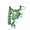
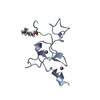
- PDB-2kfx: Structure of the N-terminal domain of human cardiac troponin C bo... -
+ Open data

- Basic information
| Entry | Database: PDB / ID: 2kfx | ||||||
|---|---|---|---|---|---|---|---|
| Title | Structure of the N-terminal domain of human cardiac troponin C bound to calcium ion and to the inhibitor W7 | ||||||
 Components Components | Troponin C, slow skeletal and cardiac muscles | ||||||
 Keywords Keywords | METAL BINDING PROTEIN / calcium regulation / striated muscle / cardiac / troponin / W7 / cardiotonic drugs / Acetylation / Calcium / Cardiomyopathy / Disease mutation / Muscle protein / Polymorphism | ||||||
| Function / homology |  Function and homology information Function and homology informationdiaphragm contraction / regulation of muscle filament sliding speed / troponin T binding / cardiac Troponin complex / troponin complex / regulation of muscle contraction / transition between fast and slow fiber / Striated Muscle Contraction / muscle filament sliding / response to metal ion ...diaphragm contraction / regulation of muscle filament sliding speed / troponin T binding / cardiac Troponin complex / troponin complex / regulation of muscle contraction / transition between fast and slow fiber / Striated Muscle Contraction / muscle filament sliding / response to metal ion / cardiac muscle cell contraction / ventricular cardiac muscle tissue morphogenesis / troponin I binding / skeletal muscle contraction / cardiac muscle contraction / sarcomere / calcium-dependent protein binding / actin filament binding / calcium ion binding / protein homodimerization activity / cytosol Similarity search - Function | ||||||
| Biological species |  Homo sapiens (human) Homo sapiens (human) | ||||||
| Method | SOLUTION NMR / molecular dynamics | ||||||
 Authors Authors | Hoffman, R.M.B. / Sykes, B.D. | ||||||
 Citation Citation | Journal: Biochemistry / Year: 2009 Title: Structure of the inhibitor W7 bound to the regulatory domain of cardiac troponin C. Authors: Hoffman, R.M. / Sykes, B.D. #1: Journal: Biochemistry / Year: 1999Title: Binding of cardiac troponin-I147-163 induces a structural opening in human cardiac troponin-C. Authors: Li, M.X. / Spyracopoulos, L. / Sykes, B.D. #2: Journal: J.Biol.Chem. / Year: 2002Title: Structure of the regulatory N-domain of human cardiac troponin C in complex with human cardiac troponin I147-163 and bepridil. Authors: Wang, X. / Li, M.X. / Sykes, B.D. | ||||||
| History |
|
- Structure visualization
| Structure viewer | Molecule: MolmilJmol/JSmol |
|---|
- Downloads & links
-Download
| PDBx/mmCIF format | 2kfx.cif.gz | 278.7 KB | Display | PDBx/mmCIF format |
|---|---|---|---|---|
| PDB format | pdb2kfx.ent.gz | 233.1 KB | Display | PDB format |
| PDBx/mmJSON format | 2kfx.json.gz | Tree view | PDBx/mmJSON format | |
| Others |  Other downloads Other downloads |
-Validation report
| Arichive directory | https://data.pdbj.org/pub/pdb/validation_reports/kf/2kfxftp://data.pdbj.org/pub/pdb/validation_reports/kf/2kfx | HTTPS FTP |
|---|
-Related structure data
| Similar structure data |
|---|


-Links
 PDBj
PDBj


- Assembly
| Deposited unit | 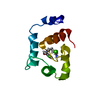
| |||||||||
|---|---|---|---|---|---|---|---|---|---|---|
| 1 |
| |||||||||
| NMR ensembles |
|
-Components
| #1: Protein | Mass: 10038.173 Da / Num. of mol.: 1 / Mutation: C35S, C84S Source method: isolated from a genetically manipulated source Source: (gene. exp.) Homo sapiens (human) / Gene: TNNC1, TNNC / Plasmid: pET / Production host:  |
|---|---|
| #2: Chemical | ChemComp-CA /   Mass: 40.078 Da / Num. of mol.: 1 / Source method: obtained synthetically / Formula: Ca Mass: 40.078 Da / Num. of mol.: 1 / Source method: obtained synthetically / Formula: Ca |
| #3: Chemical | ChemComp-WW7 / 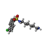  Mass: 340.868 Da / Num. of mol.: 1 / Source method: obtained synthetically / Formula: C16H21ClN2O2S Mass: 340.868 Da / Num. of mol.: 1 / Source method: obtained synthetically / Formula: C16H21ClN2O2S |
-Experimental details
-Experiment
| Experiment | Method: SOLUTION NMR Details: Structure of the N-terminal domain of human cardiac troponin C bound to the inhibitor W7 determined through isotopically edited and filtered transferred NOEs. This is based on the initial ...Details: Structure of the N-terminal domain of human cardiac troponin C bound to the inhibitor W7 determined through isotopically edited and filtered transferred NOEs. This is based on the initial coordinates of 1LXF, the intraprotein conformational restraints of 1MXL, and target geometries for a calcium-binding loop. The amine moiety of W7 is charged in this structure determination. |
|---|---|
| NMR experiment | Type: 3D-{1H,12C}-filtered-{1H,13C}-edited NOESY |
- Sample preparation
| Details | Contents: 0.8 mM [U-99% 13C; U-99% 15N] cNTnC, 4.9 mM CALCIUM ION, 0.8 mM N-(6-AMINOHEXYL)-5-CHLORO-1-NAPHTHALENESULFONAMIDE, 10 mM imidazole, 83 mM [U-99% 2H] DSS, 100% D2O Solvent system: 100% D2O | ||||||||||||||||||||||||
|---|---|---|---|---|---|---|---|---|---|---|---|---|---|---|---|---|---|---|---|---|---|---|---|---|---|
| Sample |
| ||||||||||||||||||||||||
| Sample conditions | pH: 6.75 / Temperature: 303 K |
-NMR measurement
| NMR spectrometer |
|
|---|
- Processing
| NMR software |
| ||||||||||||||||
|---|---|---|---|---|---|---|---|---|---|---|---|---|---|---|---|---|---|
| Refinement | Method: molecular dynamics / Software ordinal: 1 | ||||||||||||||||
| NMR representative | Selection criteria: lowest energy | ||||||||||||||||
| NMR ensemble | Conformer selection criteria: structures with the lowest energy Conformers calculated total number: 50 / Conformers submitted total number: 10 |
 Xplor-NIH