 Movie
Movie Controller

[English] 日本語
 Yorodumi
- PDB-2y8g: Structure of the Ran-binding domain from human RanBP3 (E352A-R353... -
+ Open data

- Basic information
| Entry | Database: PDB / ID: 2y8g | ||||||
|---|---|---|---|---|---|---|---|
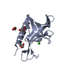
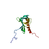
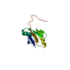
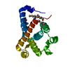
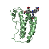
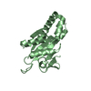
| Title | Structure of the Ran-binding domain from human RanBP3 (E352A-R353V double mutant) | ||||||
 Components Components | RAN-BINDING PROTEIN 3 | ||||||
 Keywords Keywords | PROTEIN TRANSPORT / CRM1-MEDIATED NUCLEAR EXPORT | ||||||
| Function / homology |  Function and homology information Function and homology informationR-SMAD binding / protein export from nucleus / small GTPase binding / nucleoplasm / nucleus / cytoplasm Similarity search - Function | ||||||
| Biological species |  HOMO SAPIENS (human) HOMO SAPIENS (human) | ||||||
| Method |  X-RAY DIFFRACTION / SYNCHROTRON / SIRAS / Resolution: 1.61 Å X-RAY DIFFRACTION / SYNCHROTRON / SIRAS / Resolution: 1.61 Å | ||||||
 Authors Authors | Langer, K. / Dian, C. / Rybin, V. / Muller, C.W. / Petosa, C. | ||||||
 Citation Citation | Journal: Plos One / Year: 2011 Title: Insights Into the Function of the Crm1 Cofactor Ranbp3 from the Structure of its Ran-Binding Domain Authors: Langer, K. / Dian, C. / Rybin, V. / Muller, C.W. / Petosa, C. | ||||||
| History |
|
- Structure visualization
| Structure viewer | Molecule: MolmilJmol/JSmol |
|---|
- Downloads & links
-Download
| PDBx/mmCIF format | 2y8g.cif.gz | 119.4 KB | Display | PDBx/mmCIF format |
|---|---|---|---|---|
| PDB format | pdb2y8g.ent.gz | 94.2 KB | Display | PDB format |
| PDBx/mmJSON format | 2y8g.json.gz | Tree view | PDBx/mmJSON format | |
| Others |  Other downloads Other downloads |
-Validation report
| Arichive directory | https://data.pdbj.org/pub/pdb/validation_reports/y8/2y8gftp://data.pdbj.org/pub/pdb/validation_reports/y8/2y8g | HTTPS FTP |
|---|
-Related structure data


-Links
 PDBj
PDBj- Assembly
| Deposited unit | 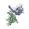
| |||||||||
|---|---|---|---|---|---|---|---|---|---|---|
| 1 | 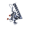
| |||||||||
| 2 | 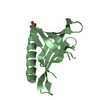
| |||||||||
| Unit cell |
| |||||||||
| Components on special symmetry positions |
|
-Components
| #1: Protein | Mass: 15431.707 Da / Num. of mol.: 2 / Fragment: RAN BINDING DOMAIN, RESIDUES 320-454 / Mutation: YES Source method: isolated from a genetically manipulated source Details: ISOFORM3 / Source: (gene. exp.) HOMO SAPIENS (human) / Plasmid: PETM11 / Production host:  #2: Chemical |   Mass: 96.063 Da / Num. of mol.: 3 / Source method: obtained synthetically / Formula: SO4 Mass: 96.063 Da / Num. of mol.: 3 / Source method: obtained synthetically / Formula: SO4#3: Water | ChemComp-HOH / |  Mass: 18.015 Da / Num. of mol.: 290 / Source method: isolated from a natural source / Formula: H2O Mass: 18.015 Da / Num. of mol.: 290 / Source method: isolated from a natural source / Formula: H2OCompound details | ENGINEERED RESIDUE IN CHAIN A, GLU 352 TO ALA ENGINEERED RESIDUE IN CHAIN A, ARG 353 TO VAL ...ENGINEERED | Sequence details | GAM REMAINING RESIDUES FROM TEV CLEAVAGE SITE | |
|---|
-Experimental details
-Experiment
| Experiment | Method: X-RAY DIFFRACTION / Number of used crystals: 2 |
|---|
- Sample preparation
| Crystal | Density Matthews: 2.8 Å3/Da / Density % sol: 56.13 % / Description: NONE |
|---|---|
| Crystal grow | pH: 7.5 / Details: 1.5 M LI2SO4, 100 MM HEPES PH 7.5 |
-Data collection
| Diffraction | Mean temperature: 100 K |
|---|---|
| Diffraction source | Source: SYNCHROTRON / Site: ESRF  / Beamline: ID23-1 / Wavelength: 1.0723 / Beamline: ID23-1 / Wavelength: 1.0723 |
| Detector | Type: ADSC CCD / Detector: CCD / Details: BENT CYLINDRICAL MIRROR |
| Radiation | Protocol: SINGLE WAVELENGTH / Monochromatic (M) / Laue (L): M / Scattering type: x-ray |
| Radiation wavelength | Wavelength: 1.0723 Å / Relative weight: 1 |
| Reflection | Resolution: 1.61→35 Å / Num. obs: 37445 / % possible obs: 94.7 % / Observed criterion σ(I): 3 / Redundancy: 10.7 % / Biso Wilson estimate: 17.69 Å2 / Rmerge(I) obs: 0.06 / Net I/σ(I): 26.7 |
| Reflection shell | Resolution: 1.61→1.7 Å / Redundancy: 9.2 % / Rmerge(I) obs: 0.53 / Mean I/σ(I) obs: 4.3 / % possible all: 81.1 |
- Processing
| Software |
| ||||||||||||||||||||||||||||||||||||||||||||||||||||||||||||||||||||||||||||||||||||||||||||||||||
|---|---|---|---|---|---|---|---|---|---|---|---|---|---|---|---|---|---|---|---|---|---|---|---|---|---|---|---|---|---|---|---|---|---|---|---|---|---|---|---|---|---|---|---|---|---|---|---|---|---|---|---|---|---|---|---|---|---|---|---|---|---|---|---|---|---|---|---|---|---|---|---|---|---|---|---|---|---|---|---|---|---|---|---|---|---|---|---|---|---|---|---|---|---|---|---|---|---|---|---|
| Refinement | Method to determine structure: SIRAS Starting model: NONE Resolution: 1.61→34.654 Å / SU ML: 0.53 / σ(F): 1.37 / Phase error: 16.56 / Stereochemistry target values: ML
| ||||||||||||||||||||||||||||||||||||||||||||||||||||||||||||||||||||||||||||||||||||||||||||||||||
| Solvent computation | Shrinkage radii: 0.9 Å / VDW probe radii: 1.11 Å / Solvent model: FLAT BULK SOLVENT MODEL / Bsol: 82.485 Å2 / ksol: 0.416 e/Å3 | ||||||||||||||||||||||||||||||||||||||||||||||||||||||||||||||||||||||||||||||||||||||||||||||||||
| Displacement parameters | Biso mean: 26.2 Å2
| ||||||||||||||||||||||||||||||||||||||||||||||||||||||||||||||||||||||||||||||||||||||||||||||||||
| Refinement step | Cycle: LAST / Resolution: 1.61→34.654 Å
| ||||||||||||||||||||||||||||||||||||||||||||||||||||||||||||||||||||||||||||||||||||||||||||||||||
| Refine LS restraints |
| ||||||||||||||||||||||||||||||||||||||||||||||||||||||||||||||||||||||||||||||||||||||||||||||||||
| LS refinement shell |
|
