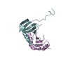
- PDB-2jql: NMR structure of the yeast Dun1 FHA domain in complex with a doub... -
+
Open data
ID or keywords:
Loading...
-
Basic information
Entry
Database: PDB / ID: 2jql
Title
NMR structure of the yeast Dun1 FHA domain in complex with a doubly phosphorylated (pT) peptide derived from Rad53 SCD1
Components
DNA damage response protein kinase DUN1
Serine/threonine-protein kinase RAD53
Keywords
CELL CYCLE / protein/phosphopeptide
Function / homology
Function and homology information
G2/M DNA damage checkpoint / Chk1/Chk2(Cds1) mediated inactivation of Cyclin B:Cdk1 complex / deoxyribonucleoside triphosphate biosynthetic process / meiotic recombination checkpoint signaling / mitotic DNA damage checkpoint signaling / dual-specificity kinase / Recruitment and ATM-mediated phosphorylation of repair and signaling proteins at DNA double strand breaks / Ubiquitin-Mediated Degradation of Phosphorylated Cdc25A / telomere maintenance in response to DNA damage / negative regulation of DNA damage checkpoint ...G2/M DNA damage checkpoint / Chk1/Chk2(Cds1) mediated inactivation of Cyclin B:Cdk1 complex / deoxyribonucleoside triphosphate biosynthetic process / meiotic recombination checkpoint signaling / mitotic DNA damage checkpoint signaling / dual-specificity kinase / Recruitment and ATM-mediated phosphorylation of repair and signaling proteins at DNA double strand breaks / Ubiquitin-Mediated Degradation of Phosphorylated Cdc25A / telomere maintenance in response to DNA damage / negative regulation of DNA damage checkpoint / replication fork processing / DNA replication origin binding / DNA replication initiation / regulation of DNA repair / protein serine/threonine/tyrosine kinase activity / DNA damage checkpoint signaling / double-strand break repair via nonhomologous end joining / intracellular protein localization / protein tyrosine kinase activity / protein kinase activity / non-specific serine/threonine protein kinase / protein serine kinase activity / DNA repair / protein serine/threonine kinase activity / ATP binding / nucleus / cytoplasm / cytosol Similarity search - Function
Serine/threonine-protein kinase Rad53 / Tumour Suppressor Smad4 - #20 / Tumour Suppressor Smad4 / Aurora kinase / Forkhead associated domain / Forkhead-associated (FHA) domain profile. / FHA domain / Forkhead-associated (FHA) domain / SMAD/FHA domain superfamily / Serine/threonine-protein kinase, active site ...Serine/threonine-protein kinase Rad53 / Tumour Suppressor Smad4 - #20 / Tumour Suppressor Smad4 / Aurora kinase / Forkhead associated domain / Forkhead-associated (FHA) domain profile. / FHA domain / Forkhead-associated (FHA) domain / SMAD/FHA domain superfamily / Serine/threonine-protein kinase, active site / Serine/Threonine protein kinases active-site signature. / Protein kinase domain / Serine/Threonine protein kinases, catalytic domain / Protein kinase, ATP binding site / Protein kinases ATP-binding region signature. / Protein kinase domain profile. / Protein kinase domain / Protein kinase-like domain superfamily / Sandwich / Mainly Beta Similarity search - Domain/homology
Mass: 1277.126 Da / Num. of mol.: 1 / Source method: obtained synthetically References: UniProt: P22216, non-specific serine/threonine protein kinase
Has protein modification
Y
-
Experimental details
-
Experiment
Experiment
Method: SOLUTION NMR
NMR experiment
Conditions-ID
Experiment-ID
Solution-ID
Type
1
1
2
3D 1H-13C NOESY
1
2
3
3D 1H-15N NOESY
1
3
3
3D HNCA
1
4
3
3DHN(CO)CA
1
5
1
3D 13C/15N-filtered (f1),13C-edited (f3) NOESY
1
6
2
3D 13C/15N-filtered (f1), 13C-edoted (f3) NOESY
1
7
2
3D 13C-edited (f1), 13C/15N-filtered (f3) NOESY
1
8
1
2D 13C/15N-filtered (f1,f2) NOESY
1
9
2
2D 13C/15N-filtered (f1,f2) NOESY
1
10
1
2D 13C/15N-filtered (f1,f2) TOCSY
1
11
1
2D 13C/15N-filtered (f1,f2) TOCSY
1
12
2
2D 13C/15N-filtered (f1,f2) COSY
-
Sample preparation
Details
Solution-ID
Contents
Solvent system
1
0.4 mM [U-13C; U-15N] protein, 0.5 mM peptide, 5 mM [U-2H] HEPES, 1 mM DTT, 150 mM NaCl, 90% H2O/10% D2O
90% H2O/10% D2O
2
0.4 mM [U-13C; U-15N] protein, 0.5 mM peptide, 5 mM [U-2H] HEPES, 150 mM NaCl, 1 mM DTT, 100% D2O
100% D2O
3
0.4 mM [U-13C; U-15N] protein, 0.5 mM peptide, 5 mM HEPES, 150 mM NaCl, 1 mM EDTA, 2 mM DTT, 90% H2O/10% D2O
90% H2O/10% D2O
Sample
Conc. (mg/ml)
Component
Isotopic labeling
Solution-ID
0.4mM
entity_1
[U-13C; U-15N]
1
0.5mM
entity_2
1
5mM
HEPES
[U-2H]
1
1mM
DTT
1
150mM
NaCl
1
0.4mM
entity_1
[U-13C; U-15N]
2
0.5mM
entity_2
2
5mM
HEPES
[U-2H]
2
150mM
NaCl
2
1mM
DTT
2
0.4mM
entity_1
[U-13C; U-15N]
3
0.5mM
entity_2
3
5mM
HEPES
3
150mM
NaCl
3
1mM
EDTA
3
2mM
DTT
3
Sample conditions
Ionic strength: 150 mM NaCl / pH: 7.5 / Pressure: ambient / Temperature: 293 K
-
NMR measurement
NMR spectrometer
Type
Manufacturer
Model
Field strength (MHz)
Spectrometer-ID
Bruker Avance
Bruker
AVANCE
800
1
Bruker DMX
Bruker
DMX
600
2
Bruker Avance
Bruker
AVANCE
500
3
-
Processing
NMR software
Name
Developer
Classification
NMRPipe
Delaglio, F. etal.
processing
NMRView
Johnson, B.A. etal.
dataanalysis
CNS
Brunger, A.T. etal.
structuresolution
CNS
Brunger, A.T. etal.
refinement
Refinement
Method: simulated annealing / Software ordinal: 1
NMR representative
Selection criteria: closest to the average
NMR ensemble
Conformer selection criteria: structures with the least restraint violations Conformers calculated total number: 100 / Conformers submitted total number: 20
+
About Yorodumi
-
News
-
Feb 9, 2022. New format data for meta-information of EMDB entries
New format data for meta-information of EMDB entries
Version 3 of the EMDB header file is now the official format.
The previous official version 1.9 will be removed from the archive.
In the structure databanks used in Yorodumi, some data are registered as the other names, "COVID-19 virus" and "2019-nCoV". Here are the details of the virus and the list of structure data.
Jan 31, 2019. EMDB accession codes are about to change! (news from PDBe EMDB page)
EMDB accession codes are about to change! (news from PDBe EMDB page)
The allocation of 4 digits for EMDB accession codes will soon come to an end. Whilst these codes will remain in use, new EMDB accession codes will include an additional digit and will expand incrementally as the available range of codes is exhausted. The current 4-digit format prefixed with “EMD-” (i.e. EMD-XXXX) will advance to a 5-digit format (i.e. EMD-XXXXX), and so on. It is currently estimated that the 4-digit codes will be depleted around Spring 2019, at which point the 5-digit format will come into force.
The EM Navigator/Yorodumi systems omit the EMD- prefix.
Related info.:Q: What is EMD? / ID/Accession-code notation in Yorodumi/EM Navigator
Yorodumi is a browser for structure data from EMDB, PDB, SASBDB, etc.
This page is also the successor to EM Navigator detail page, and also detail information page/front-end page for Omokage search.
The word "yorodu" (or yorozu) is an old Japanese word meaning "ten thousand". "mi" (miru) is to see.
Related info.:EMDB / PDB / SASBDB / Comparison of 3 databanks / Yorodumi Search / Aug 31, 2016. New EM Navigator & Yorodumi / Yorodumi Papers / Jmol/JSmol / Function and homology information / Changes in new EM Navigator and Yorodumi
 Movie
Movie Controller

 Yorodumi
 Open data
 Basic information
 Components
 Keywords
 Function and homology information
 Authors
 Citation
 Structure visualization
 Downloads & links
 Other downloads


 PDBj
PDBj


 Assembly

 HNCA
HNCA Sample preparation
 Processing