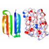
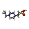
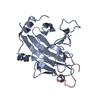
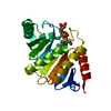
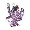
Entry Database : PDB / ID : 2jhmTitle Structure of globular heads of M-ficolin at neutral pH FICOLIN-1 Keywords / / / / / / / / Function / homology Function Domain/homology Component
/ / / / / / / / / / / / / / / / / / / / / / / / / / / / / / / / / / / / / / / / / / / / / / / / / / / / / / / / / / / / / / / / / Biological species HOMO SAPIENS (human)Method / / / Resolution : 1.52 Å Authors Garlatti, V. / Martin, L. / Gout, E. / Reiser, J.B. / Arlaud, G.J. / Thielens, N.M. / Gaboriaud, C. Journal : J.Biol.Chem. / Year : 2007Title : Structural Basis for Innate Immune Sensing by M-Ficolin and its Control by a Ph-Dependent Conformational Switch.Authors : Garlatti, V. / Martin, L. / Gout, E. / Reiser, J.B. / Fujita, T. / Arlaud, G.J. / Thielens, N.M. / Gaboriaud, C. History Deposition Feb 22, 2007 Deposition site / Processing site Revision 1.0 Oct 9, 2007 Provider / Type Revision 1.1 May 8, 2011 Group Revision 1.2 Jul 13, 2011 Group Revision 1.3 Apr 3, 2019 Group / Other / Source and taxonomyCategory / pdbx_database_proc / pdbx_database_statusItem / _pdbx_database_status.recvd_author_approvalRevision 1.4 Dec 13, 2023 Group Data collection / Database references ... Data collection / Database references / Derived calculations / Other / Refinement description Category chem_comp_atom / chem_comp_bond ... chem_comp_atom / chem_comp_bond / database_2 / pdbx_database_status / pdbx_initial_refinement_model / pdbx_struct_conn_angle / struct_conn Item _database_2.pdbx_DOI / _database_2.pdbx_database_accession ... _database_2.pdbx_DOI / _database_2.pdbx_database_accession / _pdbx_database_status.status_code_sf / _pdbx_struct_conn_angle.ptnr1_auth_comp_id / _pdbx_struct_conn_angle.ptnr1_auth_seq_id / _pdbx_struct_conn_angle.ptnr1_label_asym_id / _pdbx_struct_conn_angle.ptnr1_label_atom_id / _pdbx_struct_conn_angle.ptnr1_label_comp_id / _pdbx_struct_conn_angle.ptnr1_label_seq_id / _pdbx_struct_conn_angle.ptnr1_symmetry / _pdbx_struct_conn_angle.ptnr3_auth_comp_id / _pdbx_struct_conn_angle.ptnr3_auth_seq_id / _pdbx_struct_conn_angle.ptnr3_label_asym_id / _pdbx_struct_conn_angle.ptnr3_label_atom_id / _pdbx_struct_conn_angle.ptnr3_label_comp_id / _pdbx_struct_conn_angle.ptnr3_label_seq_id / _pdbx_struct_conn_angle.ptnr3_symmetry / _pdbx_struct_conn_angle.value / _struct_conn.pdbx_dist_value / _struct_conn.ptnr1_auth_comp_id / _struct_conn.ptnr1_auth_seq_id / _struct_conn.ptnr1_label_asym_id / _struct_conn.ptnr1_label_atom_id / _struct_conn.ptnr1_label_comp_id / _struct_conn.ptnr1_label_seq_id / _struct_conn.ptnr2_auth_comp_id / _struct_conn.ptnr2_auth_seq_id / _struct_conn.ptnr2_label_asym_id / _struct_conn.ptnr2_label_atom_id / _struct_conn.ptnr2_label_comp_id / _struct_conn.ptnr2_label_seq_id / _struct_conn.ptnr2_symmetry
Show all Show less Remark 700 SHEET THE SHEET STRUCTURE OF THIS MOLECULE IS BIFURCATED. IN ORDER TO REPRESENT THIS FEATURE IN ... SHEET THE SHEET STRUCTURE OF THIS MOLECULE IS BIFURCATED. IN ORDER TO REPRESENT THIS FEATURE IN THE SHEET RECORDS BELOW, TWO SHEETS ARE DEFINED.
 Open data
 Basic information
 Components
 Keywords
 Function and homology information
 HOMO SAPIENS (human)
 X-RAY DIFFRACTION /
  SYNCHROTRON /
  MOLECULAR REPLACEMENT / Resolution: 1.52 Å
  Authors
 Citation
 Journal: J.Biol.Chem. / Year: 2007
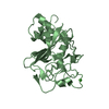
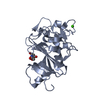
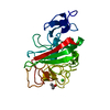
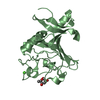
 Structure visualization
 Molmil
 Jmol/JSmol
 Downloads & links
 Download
 2jhm.cif.gz
 PDBx/mmCIF format
 pdb2jhm.ent.gz
 PDB format
 2jhm.json.gz
 PDBx/mmJSON format
 Other downloads
 2jhm_validation.pdf.gz
 wwPDB validaton report
 2jhm_full_validation.pdf.gz
 2jhm_validation.xml.gz
 2jhm_validation.cif.gz
 https://data.pdbj.org/pub/pdb/validation_reports/jh/2jhm
 ftp://data.pdbj.org/pub/pdb/validation_reports/jh/2jhm


 Links
Links Assembly
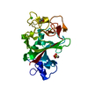
 Components
 HOMO SAPIENS (human) / Cell line (production host): High Five / Production host:
  TRICHOPLUSIA NI (cabbage looper) / References: UniProt: O00602
 X-RAY DIFFRACTION / Number of used crystals: 1
  Sample preparation
 SYNCHROTRON / Site:
  ESRF
ESRF  / Beamline: ID14-2 / Wavelength: 0.933
  Processing
 MOLECULAR REPLACEMENT
 Movie
Movie Controller




 PDBj
PDBj


