 Movie
Movie Controller

[English] 日本語
 Yorodumi
- PDB-2jex: Transcription activator structure reveals redox control of a repl... -
+ Open data

- Basic information
| Entry | Database: PDB / ID: 2jex | ||||||
|---|---|---|---|---|---|---|---|
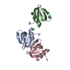
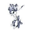
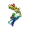
| Title | Transcription activator structure reveals redox control of a replication initiation reaction | ||||||
 Components Components | REGULATORY PROTEIN E2 | ||||||
 Keywords Keywords | TRANSCRIPTION / NUCLEAR PROTEIN / DNA REPLICATION / PHOSPHORYLATION / TRANSCRIPTION REGULATION / VIRAL TRANSCRIPTION FACTOR / BOVINE PAPILLOMAVIRUS / REPLICATION INITIATION / EARLY PROTEIN / REDOX CONTROL / E1 / E2 / OXIDATION / ACTIVATOR / REPRESSOR / DNA-BINDING | ||||||
| Function / homology |  Function and homology information Function and homology informationviral DNA genome replication / regulation of DNA replication / DNA replication / DNA-binding transcription factor activity / nucleotide binding / DNA-templated transcription / host cell nucleus / DNA binding Similarity search - Function | ||||||
| Biological species |  BOVINE PAPILLOMAVIRUS TYPE 1 BOVINE PAPILLOMAVIRUS TYPE 1 | ||||||
| Method |  X-RAY DIFFRACTION / SYNCHROTRON / MOLECULAR REPLACEMENT / Resolution: 2.35 Å X-RAY DIFFRACTION / SYNCHROTRON / MOLECULAR REPLACEMENT / Resolution: 2.35 Å | ||||||
 Authors Authors | Sanders, C.M. / Sizov, D. / Seavers, P.R. / Ortiz-Lombardia, M. / Antson, A.A. | ||||||
 Citation Citation | Journal: Nucleic Acids Res. / Year: 2007 Title: Transcription Activator Structure Reveals Redox Control of a Replication Initiation Reaction. Authors: Sanders, C.M. / Sizov, D. / Seavers, P.R. / Ortiz-Lombardia, M. / Antson, A.A. | ||||||
| History |
|
- Structure visualization
| Structure viewer | Molecule: MolmilJmol/JSmol |
|---|
- Downloads & links
-Download
| PDBx/mmCIF format | 2jex.cif.gz | 55.5 KB | Display | PDBx/mmCIF format |
|---|---|---|---|---|
| PDB format | pdb2jex.ent.gz | 39 KB | Display | PDB format |
| PDBx/mmJSON format | 2jex.json.gz | Tree view | PDBx/mmJSON format | |
| Others |  Other downloads Other downloads |
-Validation report
| Arichive directory | https://data.pdbj.org/pub/pdb/validation_reports/je/2jexftp://data.pdbj.org/pub/pdb/validation_reports/je/2jex | HTTPS FTP |
|---|
-Related structure data
| Related structure data |  2jeuSC S: Starting model for refinement C: citing same article ( |
|---|---|
| Similar structure data |


-Links
 PDBj
PDBj- Assembly
| Deposited unit | 
| ||||||||
|---|---|---|---|---|---|---|---|---|---|
| 1 |
| ||||||||
| Unit cell |
|
-Components
| #1: Protein | Mass: 23820.590 Da / Num. of mol.: 1 Fragment: N-TERMINAL TRANS-ACTIVATION DOMAIN (TAD), RESIDUES 1-209 Mutation: YES Source method: isolated from a genetically manipulated source Source: (gene. exp.) BOVINE PAPILLOMAVIRUS TYPE 1 / Production host:  |
|---|---|
| #2: Water | ChemComp-HOH /  Mass: 18.015 Da / Num. of mol.: 115 / Source method: isolated from a natural source / Formula: H2O Mass: 18.015 Da / Num. of mol.: 115 / Source method: isolated from a natural source / Formula: H2O |
| Compound details | ENGINEERED |
-Experimental details
-Experiment
| Experiment | Method: X-RAY DIFFRACTION / Number of used crystals: 1 |
|---|
- Sample preparation
| Crystal | Density Matthews: 4.01 Å3/Da / Density % sol: 69.06 % |
|---|---|
| Crystal grow | Method: vapor diffusion, hanging drop / pH: 8.5 Details: PROTEIN WAS CRYSTALLISED FROM 0.1 M TRIS-HCL PH 8.5, 0.3 M NACL, 2MM DTT AND 18-22% TERTIARY BUTANOL, USING THE HANGING DROP METHOD OF VAPOUR DIFFUSION. |
-Data collection
| Diffraction | Mean temperature: 120 K |
|---|---|
| Diffraction source | Source: SYNCHROTRON / Site: ESRF  / Beamline: BM14 / Wavelength: 0.919 / Beamline: BM14 / Wavelength: 0.919 |
| Detector | Type: MARRESEARCH / Detector: CCD |
| Radiation | Protocol: SINGLE WAVELENGTH / Monochromatic (M) / Laue (L): M / Scattering type: x-ray |
| Radiation wavelength | Wavelength: 0.919 Å / Relative weight: 1 |
| Reflection | Resolution: 2.35→25 Å / Num. obs: 14352 / % possible obs: 93.3 % / Observed criterion σ(I): 3 / Redundancy: 1.9 % / Rmerge(I) obs: 0.07 / Net I/σ(I): 10.7 |
| Reflection shell | Resolution: 2.35→2.43 Å / Redundancy: 1.8 % / Rmerge(I) obs: 0.32 / Mean I/σ(I) obs: 2.5 / % possible all: 90.2 |
- Processing
| Software |
| ||||||||||||||||||||||||||||||||||||||||||||||||||||||||||||||||||||||||||||||||||||||||||||||||||||||||||||||||||||||||||||||||||||||||||||||||||||||||||||||||||||||||||||||||||||||
|---|---|---|---|---|---|---|---|---|---|---|---|---|---|---|---|---|---|---|---|---|---|---|---|---|---|---|---|---|---|---|---|---|---|---|---|---|---|---|---|---|---|---|---|---|---|---|---|---|---|---|---|---|---|---|---|---|---|---|---|---|---|---|---|---|---|---|---|---|---|---|---|---|---|---|---|---|---|---|---|---|---|---|---|---|---|---|---|---|---|---|---|---|---|---|---|---|---|---|---|---|---|---|---|---|---|---|---|---|---|---|---|---|---|---|---|---|---|---|---|---|---|---|---|---|---|---|---|---|---|---|---|---|---|---|---|---|---|---|---|---|---|---|---|---|---|---|---|---|---|---|---|---|---|---|---|---|---|---|---|---|---|---|---|---|---|---|---|---|---|---|---|---|---|---|---|---|---|---|---|---|---|---|---|
| Refinement | Method to determine structure: MOLECULAR REPLACEMENT Starting model: PDB ENTRY 2JEU Resolution: 2.35→22.62 Å / Cor.coef. Fo:Fc: 0.948 / Cor.coef. Fo:Fc free: 0.936 / SU B: 15.331 / SU ML: 0.188 / TLS residual ADP flag: LIKELY RESIDUAL / Cross valid method: THROUGHOUT / ESU R: 0.251 / ESU R Free: 0.204 / Stereochemistry target values: MAXIMUM LIKELIHOOD
| ||||||||||||||||||||||||||||||||||||||||||||||||||||||||||||||||||||||||||||||||||||||||||||||||||||||||||||||||||||||||||||||||||||||||||||||||||||||||||||||||||||||||||||||||||||||
| Solvent computation | Ion probe radii: 0.8 Å / Shrinkage radii: 0.8 Å / VDW probe radii: 1.2 Å / Solvent model: BABINET MODEL WITH MASK | ||||||||||||||||||||||||||||||||||||||||||||||||||||||||||||||||||||||||||||||||||||||||||||||||||||||||||||||||||||||||||||||||||||||||||||||||||||||||||||||||||||||||||||||||||||||
| Displacement parameters | Biso mean: 72.2 Å2
| ||||||||||||||||||||||||||||||||||||||||||||||||||||||||||||||||||||||||||||||||||||||||||||||||||||||||||||||||||||||||||||||||||||||||||||||||||||||||||||||||||||||||||||||||||||||
| Refinement step | Cycle: LAST / Resolution: 2.35→22.62 Å
| ||||||||||||||||||||||||||||||||||||||||||||||||||||||||||||||||||||||||||||||||||||||||||||||||||||||||||||||||||||||||||||||||||||||||||||||||||||||||||||||||||||||||||||||||||||||
| Refine LS restraints |
|