 Movie
Movie Controller


+ Open data

- Basic information
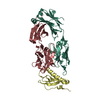
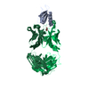
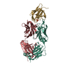
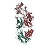
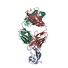
| Entry | Database: PDB / ID: 2jel | |||||||||
|---|---|---|---|---|---|---|---|---|---|---|
| Title | JEL42 FAB/HPR COMPLEX | |||||||||
 Components Components |
| |||||||||
 Keywords Keywords | COMPLEX (ANTIBODY/ANTIGEN) / ANTIBODY-PROTEIN COMPLEX / HISTIDINE-CONTAINING PROTEIN / MOLECULAR RECOGNITION / COMPLEX (ANTIBODY-ANTIGEN) / COMPLEX (ANTIBODY-ANTIGEN) complex | |||||||||
| Function / homology |  Function and homology information Function and homology informationphosphotransferase activity, nitrogenous group as acceptor / antisigma factor binding / regulation of carbon utilization / peptidyl-histidine phosphorylation / positive regulation of glycogen catabolic process / phosphoenolpyruvate-dependent sugar phosphotransferase system / enzyme regulator activity / enzyme inhibitor activity / enzyme activator activity / cytosol Similarity search - Function | |||||||||
| Biological species |   | |||||||||
| Method |  X-RAY DIFFRACTION / MOLECULAR REPLACEMENT / Resolution: 2.5 Å X-RAY DIFFRACTION / MOLECULAR REPLACEMENT / Resolution: 2.5 Å | |||||||||
 Authors Authors | Prasad, L. / Waygood, E.B. / Lee, J.S. / Delbaere, L.T.J. | |||||||||
 Citation Citation | Journal: J.Mol.Biol. / Year: 1998 Title: The 2.5 A resolution structure of the jel42 Fab fragment/HPr complex Authors: Prasad, L. / Waygood, E.B. / Lee, J.S. / Delbaere, L.T.J. #1: Journal: J.Biol.Chem. / Year: 1993Title: Evaluation of Mutagenesis for Epitope Mapping. Structure of an Antibody-Protein Antigen Complex Authors: Prasad, L. / Sharma, S. / Vandonselaar, M. / Quail, J.W. / Lee, J.S. / Waygood, E.B. / Wilson, K.S. / Dauter, Z. / Delbaere, L.T. #2: Journal: Proc.Natl.Acad.Sci.USA / Year: 1991Title: Epitope Mapping by Mutagenesis Distinguishes between the Two Tertiary Structures of the Histidine-Containing Protein Hpr Authors: Sharma, S. / Georges, F. / Delbaere, L.T. / Lee, J.S. / Klevit, R.E. / Waygood, E.B. #3: Journal: J.Biol.Chem. / Year: 1989Title: Crystallization of the Complex of a Monoclonal Fab Fragment with the Histidine-Containing Protein of the Phosphoenolpyruvate: Sugar Phosphotransferase System of Escherichia Coli Authors: Delbaere, L.T. / Vandonselaar, M. / Quail, J.W. / Waygood, E.B. / Lee, J.S. #4: Journal: J.Biol.Chem. / Year: 1988Title: Structure Determination of a Monoclonal Fab Fragment Specific for Histidine-Containing Protein of the Phosphoenolpyruvate: Sugar Phosphotransferase System of Escherichia Coli Authors: Prasad, L. / Vandonselaar, M. / Lee, J.S. / Delbaere, L.T. | |||||||||
| History |
|
- Structure visualization
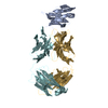
| Structure viewer | Molecule: MolmilJmol/JSmol |
|---|
- Downloads & links
-Download
| PDBx/mmCIF format | 2jel.cif.gz | 112.5 KB | Display | PDBx/mmCIF format |
|---|---|---|---|---|
| PDB format | pdb2jel.ent.gz | 86 KB | Display | PDB format |
| PDBx/mmJSON format | 2jel.json.gz | Tree view | PDBx/mmJSON format | |
| Others |  Other downloads Other downloads |
-Validation report
| Arichive directory | https://data.pdbj.org/pub/pdb/validation_reports/je/2jelftp://data.pdbj.org/pub/pdb/validation_reports/je/2jel | HTTPS FTP |
|---|
-Related structure data
| Similar structure data |
|---|


-Links
 PDBj
PDBj

- Assembly
| Deposited unit | 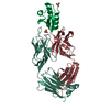
| ||||||||
|---|---|---|---|---|---|---|---|---|---|
| 1 |
| ||||||||
| Unit cell |
|
-Components
| #1: Antibody | Mass: 23860.471 Da / Num. of mol.: 1 / Source method: isolated from a natural source / Source: (natural) | ||||||
|---|---|---|---|---|---|---|---|
| #2: Antibody | Mass: 23094.049 Da / Num. of mol.: 1 / Source method: isolated from a natural source / Source: (natural) | ||||||
| #3: Protein | Mass: 9129.332 Da / Num. of mol.: 1 / Source method: isolated from a natural source / Source: (natural) | ||||||
| #4: Chemical |   Mass: 96.063 Da / Num. of mol.: 2 / Source method: obtained synthetically / Formula: SO4 Mass: 96.063 Da / Num. of mol.: 2 / Source method: obtained synthetically / Formula: SO4#5: Water | ChemComp-HOH / |  Mass: 18.015 Da / Num. of mol.: 67 / Source method: isolated from a natural source / Formula: H2O Mass: 18.015 Da / Num. of mol.: 67 / Source method: isolated from a natural source / Formula: H2OHas protein modification | Y | Sequence details | RESIDUES OF THE FAB FRAGMENT ARE NUMBERED ACCORDING TO KABAT. | |
-Experimental details
-Experiment
| Experiment | Method: X-RAY DIFFRACTION / Number of used crystals: 1 |
|---|
- Sample preparation
| Crystal | Density Matthews: 2.96 Å3/Da / Density % sol: 57 % | |||||||||||||||||||||||||
|---|---|---|---|---|---|---|---|---|---|---|---|---|---|---|---|---|---|---|---|---|---|---|---|---|---|---|
| Crystal grow | pH: 5.8 / Details: pH 5.8 | |||||||||||||||||||||||||
| Crystal grow | *PLUS Method: vapor diffusion, sitting dropDetails: the molar ratio of Fab fragment to HPr was approximately 1:2 | |||||||||||||||||||||||||
| Components of the solutions | *PLUS
|
-Data collection
| Diffraction | Mean temperature: 289 K |
|---|---|
| Diffraction source | Source: ROTATING ANODE / Type: ENRAF-NONIUS FR571 / Wavelength: 1.5418 |
| Detector | Type: ENRAF-NONIUS FAST / Detector: DIFFRACTOMETER / Date: Apr 1, 1992 / Details: COLLIMATOR |
| Radiation | Monochromator: GRAPHITE(002) / Monochromatic (M) / Laue (L): M / Scattering type: x-ray |
| Radiation wavelength | Wavelength: 1.5418 Å / Relative weight: 1 |
| Reflection | Resolution: 2.5→35 Å / Num. obs: 22067 / % possible obs: 96.6 % / Observed criterion σ(I): 5 / Redundancy: 2.7 % / Rsym value: 0.046 / Net I/σ(I): 11 |
| Reflection shell | Resolution: 2.5→2.56 Å / Redundancy: 2.5 % / Mean I/σ(I) obs: 3.5 / Rsym value: 0.19 / % possible all: 93 |
| Reflection | *PLUS Num. measured all: 47817 / Rmerge(I) obs: 0.046 |
| Reflection shell | *PLUS % possible obs: 93 % / Num. unique obs: 1043 / Num. measured obs: 2784 / Rmerge(I) obs: 0.19 |
- Processing
| Software |
| ||||||||||||||||||||||||||||||||||||||||||||||||||||||||||||||||||||||||||||||||
|---|---|---|---|---|---|---|---|---|---|---|---|---|---|---|---|---|---|---|---|---|---|---|---|---|---|---|---|---|---|---|---|---|---|---|---|---|---|---|---|---|---|---|---|---|---|---|---|---|---|---|---|---|---|---|---|---|---|---|---|---|---|---|---|---|---|---|---|---|---|---|---|---|---|---|---|---|---|---|---|---|---|
| Refinement | Method to determine structure: MOLECULAR REPLACEMENT Starting model: GLOOP2 FAB Resolution: 2.5→6 Å / Rfactor Rfree error: 0.01 / Data cutoff high absF: 10000000 / Data cutoff low absF: 0 / Isotropic thermal model: INDIVIDUAL ISOTROPIC / Cross valid method: THROUGHOUT / σ(F): 0
| ||||||||||||||||||||||||||||||||||||||||||||||||||||||||||||||||||||||||||||||||
| Displacement parameters | Biso mean: 36 Å2 | ||||||||||||||||||||||||||||||||||||||||||||||||||||||||||||||||||||||||||||||||
| Refine analyze |
| ||||||||||||||||||||||||||||||||||||||||||||||||||||||||||||||||||||||||||||||||
| Refinement step | Cycle: LAST / Resolution: 2.5→6 Å
| ||||||||||||||||||||||||||||||||||||||||||||||||||||||||||||||||||||||||||||||||
| Refine LS restraints |
| ||||||||||||||||||||||||||||||||||||||||||||||||||||||||||||||||||||||||||||||||
| LS refinement shell | Resolution: 2.5→2.56 Å / Rfactor Rfree error: 0.036 / Total num. of bins used: 8
| ||||||||||||||||||||||||||||||||||||||||||||||||||||||||||||||||||||||||||||||||
| Xplor file |
| ||||||||||||||||||||||||||||||||||||||||||||||||||||||||||||||||||||||||||||||||
| Software | *PLUS Name: X-PLOR / Version: 3.1 / Classification: refinement | ||||||||||||||||||||||||||||||||||||||||||||||||||||||||||||||||||||||||||||||||
| Refinement | *PLUS | ||||||||||||||||||||||||||||||||||||||||||||||||||||||||||||||||||||||||||||||||
| Solvent computation | *PLUS | ||||||||||||||||||||||||||||||||||||||||||||||||||||||||||||||||||||||||||||||||
| Displacement parameters | *PLUS | ||||||||||||||||||||||||||||||||||||||||||||||||||||||||||||||||||||||||||||||||
| Refine LS restraints | *PLUS
| ||||||||||||||||||||||||||||||||||||||||||||||||||||||||||||||||||||||||||||||||
| LS refinement shell | *PLUS Rfactor obs: 0.34 |
