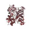
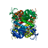
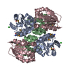
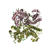
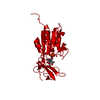
Entry Database : PDB / ID : 2ja1Title Thymidine kinase from B. cereus with TTP bound as phosphate donor. THYMIDINE KINASE Keywords / / / / / / Function / homology Function Domain/homology Component
/ / / / / / / / / / / / / / / / / / / / / / / / / / Biological species BACILLUS CEREUS (bacteria)Method / / / Resolution : 2.8 Å Authors Kosinska, U. / Carnrot, C. / Sandrini, M.P.B. / Clausen, A.R. / Wang, L. / Piskur, J. / Eriksson, S. / Eklund, H. Journal : FEBS J. / Year : 2007Title : Structural Studies of Thymidine Kinases from Bacillus Anthracis and Bacillus Cereus Provide Insights Into Quaternary Structure and Conformational Changes Upon Substrate BindingAuthors : Kosinska, U. / Carnrot, C. / Sandrini, M.P.B. / Clausen, A.R. / Wang, L. / Piskur, J. / Eriksson, S. / Eklund, H. History Deposition Nov 17, 2006 Deposition site / Processing site Revision 1.0 Jan 23, 2007 Provider / Type Revision 1.1 Jul 13, 2011 Group / Version format complianceRevision 1.2 Sep 4, 2019 Group / Category / reflns_shellItem _reflns.pdbx_Rmerge_I_obs / _reflns.pdbx_Rrim_I_all ... _reflns.pdbx_Rmerge_I_obs / _reflns.pdbx_Rrim_I_all / _reflns_shell.Rmerge_I_obs / _reflns_shell.pdbx_Rrim_I_all Revision 1.3 Dec 13, 2023 Group Data collection / Database references ... Data collection / Database references / Derived calculations / Other / Refinement description Category chem_comp_atom / chem_comp_bond ... chem_comp_atom / chem_comp_bond / database_2 / pdbx_database_status / pdbx_initial_refinement_model / struct_conn Item _database_2.pdbx_DOI / _database_2.pdbx_database_accession ... _database_2.pdbx_DOI / _database_2.pdbx_database_accession / _pdbx_database_status.status_code_sf / _struct_conn.ptnr1_auth_comp_id / _struct_conn.ptnr1_auth_seq_id / _struct_conn.ptnr1_label_asym_id / _struct_conn.ptnr1_label_atom_id / _struct_conn.ptnr1_label_comp_id / _struct_conn.ptnr1_label_seq_id / _struct_conn.ptnr2_auth_comp_id / _struct_conn.ptnr2_auth_seq_id / _struct_conn.ptnr2_label_asym_id / _struct_conn.ptnr2_label_atom_id / _struct_conn.ptnr2_label_comp_id / _struct_conn.ptnr2_label_seq_id
Show all Show less Remark 700 SHEET DETERMINATION METHOD: AUTHOR PROVIDED.
 Movie
Movie Controller

 Yorodumi
 Open data
 Basic information
 Components
 Keywords
 Function and homology information
 X-RAY DIFFRACTION /
  Authors
 Citation
 Structure visualization
 Downloads & links
 Other downloads
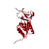


 PDBj
PDBj
 Assembly
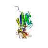

 Mass: 65.409 Da / Num. of mol.: 1 / Source method: obtained synthetically / Formula: Zn
 Mass: 482.168 Da / Num. of mol.: 1 / Source method: obtained synthetically / Formula: C10H17N2O14P3
 Mass: 118.174 Da / Num. of mol.: 1 / Source method: obtained synthetically / Formula: C6H14O2 / Comment: precipitant*YM
 Mass: 18.015 Da / Num. of mol.: 20 / Source method: isolated from a natural source / Formula: H2O
 Sample preparation
 / Beamline: ID14-2 / Wavelength: 0.933
  Processing