 Movie
Movie Controller

[English] 日本語
 Yorodumi
- PDB-6oww: Crystal structure of a Human Cardiac Calsequestrin Filament Compl... -
+ Open data

- Basic information
| Entry | Database: PDB / ID: 6oww | |||||||||||||||
|---|---|---|---|---|---|---|---|---|---|---|---|---|---|---|---|---|
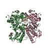
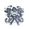
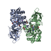
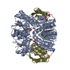
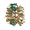
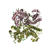
| Title | Crystal structure of a Human Cardiac Calsequestrin Filament Complexed with Ytterbium | |||||||||||||||
 Components Components | Calsequestrin-2 | |||||||||||||||
 Keywords Keywords | METAL BINDING PROTEIN / Calsequestrin / Calcium-Binding Proteins / Sarcoplasmic Reticulum Proteins | |||||||||||||||
| Function / homology |  Function and homology information Function and homology informationcalcium ion sequestering activity / regulation of membrane repolarization during ventricular cardiac muscle cell action potential / regulation of cell communication by electrical coupling / junctional sarcoplasmic reticulum membrane / Purkinje myocyte to ventricular cardiac muscle cell signaling / : / sarcoplasmic reticulum lumen / regulation of membrane repolarization / cellular response to caffeine / negative regulation of potassium ion transport ...calcium ion sequestering activity / regulation of membrane repolarization during ventricular cardiac muscle cell action potential / regulation of cell communication by electrical coupling / junctional sarcoplasmic reticulum membrane / Purkinje myocyte to ventricular cardiac muscle cell signaling / : / sarcoplasmic reticulum lumen / regulation of membrane repolarization / cellular response to caffeine / negative regulation of potassium ion transport / protein polymerization / detection of calcium ion / striated muscle contraction / regulation of release of sequestered calcium ion into cytosol by sarcoplasmic reticulum / Ion homeostasis / cardiac muscle contraction / regulation of cardiac muscle contraction by regulation of the release of sequestered calcium ion / sarcoplasmic reticulum membrane / calcium channel complex / regulation of heart rate / sarcoplasmic reticulum / Stimuli-sensing channels / Z disc / intracellular calcium ion homeostasis / calcium-dependent protein binding / calcium ion binding / protein homodimerization activity / cytoplasm Similarity search - Function | |||||||||||||||
| Biological species |  Homo sapiens (human) Homo sapiens (human) | |||||||||||||||
| Method |  X-RAY DIFFRACTION / SYNCHROTRON / MOLECULAR REPLACEMENT / Resolution: 3.84 Å X-RAY DIFFRACTION / SYNCHROTRON / MOLECULAR REPLACEMENT / Resolution: 3.84 Å | |||||||||||||||
 Authors Authors | Titus, E.W. / Deiter, F.H. / Shi, C. / Jura, N. / Deo, R.C. | |||||||||||||||
| Funding support |  United States, 4items United States, 4items
| |||||||||||||||
 Citation Citation | Journal: Nat.Struct.Mol.Biol. / Year: 2020 Title: The structure of a calsequestrin filament reveals mechanisms of familial arrhythmia. Authors: Titus, E.W. / Deiter, F.H. / Shi, C. / Wojciak, J. / Scheinman, M. / Jura, N. / Deo, R.C. | |||||||||||||||
| History |
|
- Structure visualization
| Structure viewer | Molecule: MolmilJmol/JSmol |
|---|
- Downloads & links
-Download
| PDBx/mmCIF format | 6oww.cif.gz | 543.4 KB | Display | PDBx/mmCIF format |
|---|---|---|---|---|
| PDB format | pdb6oww.ent.gz | 444.7 KB | Display | PDB format |
| PDBx/mmJSON format | 6oww.json.gz | Tree view | PDBx/mmJSON format | |
| Others |  Other downloads Other downloads |
-Validation report
| Arichive directory | https://data.pdbj.org/pub/pdb/validation_reports/ow/6owwftp://data.pdbj.org/pub/pdb/validation_reports/ow/6oww | HTTPS FTP |
|---|
-Related structure data
| Related structure data |  6owvSC S: Starting model for refinement C: citing same article ( |
|---|---|
| Similar structure data | |
| Experimental dataset #1 | Data reference: 10.5281/zenodo.2943248 / Data set type: diffraction image data |


-Links
 PDBj
PDBj


- Assembly
| Deposited unit | 
| ||||||||||
|---|---|---|---|---|---|---|---|---|---|---|---|
| 1 | 
| ||||||||||
| 2 | 
| ||||||||||
| 3 | 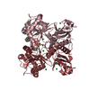
| ||||||||||
| 4 | 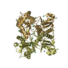
| ||||||||||
| Unit cell |
|
-Components
| #1: Protein | Mass: 44968.637 Da / Num. of mol.: 8 Source method: isolated from a genetically manipulated source Source: (gene. exp.) Homo sapiens (human) / Gene: CASQ2 / Production host:  #2: Chemical | ChemComp-YB /   Mass: 173.040 Da / Num. of mol.: 63 / Source method: obtained synthetically / Formula: Yb / Feature type: SUBJECT OF INVESTIGATION Mass: 173.040 Da / Num. of mol.: 63 / Source method: obtained synthetically / Formula: Yb / Feature type: SUBJECT OF INVESTIGATION#3: Chemical | ChemComp-SO4 /   Mass: 96.063 Da / Num. of mol.: 5 / Source method: obtained synthetically / Formula: SO4 Mass: 96.063 Da / Num. of mol.: 5 / Source method: obtained synthetically / Formula: SO4Has ligand of interest | Y | |
|---|
-Experimental details
-Experiment
| Experiment | Method: X-RAY DIFFRACTION / Number of used crystals: 1 |
|---|
- Sample preparation
| Crystal | Density Matthews: 2.2 Å3/Da / Density % sol: 44.19 % |
|---|---|
| Crystal grow | Temperature: 298 K / Method: vapor diffusion, hanging drop / Details: PEG 4000, lithium sulfate / Temp details: Room temperature |
-Data collection
| Diffraction | Mean temperature: 100 K / Ambient temp details: cryo / Serial crystal experiment: N | ||||||||||||||||||||||||
|---|---|---|---|---|---|---|---|---|---|---|---|---|---|---|---|---|---|---|---|---|---|---|---|---|---|
| Diffraction source | Source: SYNCHROTRON / Site: ALS / Beamline: 8.3.1 / Wavelength: 1.3857 Å | ||||||||||||||||||||||||
| Detector | Type: DECTRIS PILATUS3 S 6M / Detector: PIXEL / Date: Aug 17, 2017 | ||||||||||||||||||||||||
| Radiation | Protocol: SINGLE WAVELENGTH / Monochromatic (M) / Laue (L): M / Scattering type: x-ray | ||||||||||||||||||||||||
| Radiation wavelength | Wavelength: 1.3857 Å / Relative weight: 1 | ||||||||||||||||||||||||
| Reflection | Resolution: 3.84→214.34 Å / Num. obs: 29912 / % possible obs: 98.9 % / Redundancy: 11.2 % / Biso Wilson estimate: 59.129 Å2 / Rpim(I) all: 0.162 / Rrim(I) all: 0.548 / Net I/σ(I): 4.5 | ||||||||||||||||||||||||
| Reflection shell | Diffraction-ID: 1
|
- Processing
| Software |
| ||||||||||||||||||||||||||||||||||||||||||||||||||||||||||||||||||||||||||||||||||||||||||||||||||||||||||||||||||||||||||||||
|---|---|---|---|---|---|---|---|---|---|---|---|---|---|---|---|---|---|---|---|---|---|---|---|---|---|---|---|---|---|---|---|---|---|---|---|---|---|---|---|---|---|---|---|---|---|---|---|---|---|---|---|---|---|---|---|---|---|---|---|---|---|---|---|---|---|---|---|---|---|---|---|---|---|---|---|---|---|---|---|---|---|---|---|---|---|---|---|---|---|---|---|---|---|---|---|---|---|---|---|---|---|---|---|---|---|---|---|---|---|---|---|---|---|---|---|---|---|---|---|---|---|---|---|---|---|---|---|
| Refinement | Method to determine structure: MOLECULAR REPLACEMENT Starting model: 6OWV Resolution: 3.84→214.34 Å / SU ML: 0.67 / Cross valid method: THROUGHOUT / σ(F): 1.97 / Phase error: 37.13 / Stereochemistry target values: ML
| ||||||||||||||||||||||||||||||||||||||||||||||||||||||||||||||||||||||||||||||||||||||||||||||||||||||||||||||||||||||||||||||
| Solvent computation | Shrinkage radii: 0.8 Å / VDW probe radii: 1 Å / Solvent model: FLAT BULK SOLVENT MODEL | ||||||||||||||||||||||||||||||||||||||||||||||||||||||||||||||||||||||||||||||||||||||||||||||||||||||||||||||||||||||||||||||
| Displacement parameters | Biso max: 347.97 Å2 / Biso mean: 98.7266 Å2 / Biso min: 41.16 Å2 | ||||||||||||||||||||||||||||||||||||||||||||||||||||||||||||||||||||||||||||||||||||||||||||||||||||||||||||||||||||||||||||||
| Refinement step | Cycle: final / Resolution: 3.84→214.34 Å
| ||||||||||||||||||||||||||||||||||||||||||||||||||||||||||||||||||||||||||||||||||||||||||||||||||||||||||||||||||||||||||||||
| LS refinement shell | Refine-ID: X-RAY DIFFRACTION / Rfactor Rfree error: 0
|
