 Movie
Movie Controller

[English] 日本語
 Yorodumi
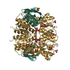
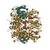
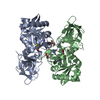
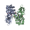
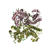
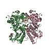
- PDB-2fep: Structure of truncated CcpA in complex with P-Ser-HPr and Sulfate ions -
+ Open data

- Basic information
| Entry | Database: PDB / ID: 2fep | ||||||
|---|---|---|---|---|---|---|---|
| Title | Structure of truncated CcpA in complex with P-Ser-HPr and Sulfate ions | ||||||
 Components Components |
| ||||||
 Keywords Keywords | TRANSCRIPTION / CcpA / HPr / transcriptional regulator | ||||||
| Function / homology |  Function and homology information Function and homology informationregulation of carbohydrate utilization / phosphoenolpyruvate-dependent sugar phosphotransferase system / DNA-binding transcription repressor activity / DNA-binding transcription activator activity / protein-DNA complex / transcription cis-regulatory region binding / DNA-binding transcription factor activity / negative regulation of DNA-templated transcription / regulation of DNA-templated transcription / positive regulation of DNA-templated transcription / cytoplasm Similarity search - Function | ||||||
| Biological species |  | ||||||
| Method |  X-RAY DIFFRACTION / SYNCHROTRON / MOLECULAR REPLACEMENT / Resolution: 2.45 Å X-RAY DIFFRACTION / SYNCHROTRON / MOLECULAR REPLACEMENT / Resolution: 2.45 Å | ||||||
 Authors Authors | Chaptal, V. / Gueguen-Chaignon, V. / Poncet, S. / Lecampion, C. / Meyer, P. / Deutscher, J. / Galinier, A. / Nessler, S. | ||||||
 Citation Citation | Journal: Proteins / Year: 2006 Title: Structural analysis of B. subtilis CcpA effector binding site. Authors: Chaptal, V. / Gueguen-Chaignon, V. / Poncet, S. / Lecampion, C. / Meyer, P. / Deutscher, J. / Galinier, A. / Nessler, S. / Morera, S. | ||||||
| History |
|
- Structure visualization
| Structure viewer | Molecule: MolmilJmol/JSmol |
|---|
- Downloads & links
-Download
| PDBx/mmCIF format | 2fep.cif.gz | 84.3 KB | Display | PDBx/mmCIF format |
|---|---|---|---|---|
| PDB format | pdb2fep.ent.gz | 62.4 KB | Display | PDB format |
| PDBx/mmJSON format | 2fep.json.gz | Tree view | PDBx/mmJSON format | |
| Others |  Other downloads Other downloads |
-Validation report
| Arichive directory | https://data.pdbj.org/pub/pdb/validation_reports/fe/2fepftp://data.pdbj.org/pub/pdb/validation_reports/fe/2fep | HTTPS FTP |
|---|
-Related structure data
| Related structure data |  1rzrS S: Starting model for refinement |
|---|---|
| Similar structure data |


-Links
 PDBj
PDBj


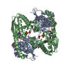
- Assembly
| Deposited unit | 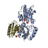
| ||||||||
|---|---|---|---|---|---|---|---|---|---|
| 1 | 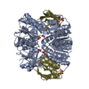
| ||||||||
| Unit cell |
| ||||||||
| Details | The biological assembly is a dimer generated from the monomer in the asymetric unit by a two fold crystallographic axis |
-Components
| #1: Protein | Mass: 32183.639 Da / Num. of mol.: 1 / Fragment: residues 61-333 Source method: isolated from a genetically manipulated source Source: (gene. exp.) | ||||
|---|---|---|---|---|---|
| #2: Protein | Mass: 9278.313 Da / Num. of mol.: 1 Source method: isolated from a genetically manipulated source Source: (gene. exp.) | ||||
| #3: Chemical |   Mass: 96.063 Da / Num. of mol.: 3 / Source method: obtained synthetically / Formula: SO4 Mass: 96.063 Da / Num. of mol.: 3 / Source method: obtained synthetically / Formula: SO4#4: Water | ChemComp-HOH / |  Mass: 18.015 Da / Num. of mol.: 106 / Source method: isolated from a natural source / Formula: H2O Mass: 18.015 Da / Num. of mol.: 106 / Source method: isolated from a natural source / Formula: H2OHas protein modification | Y | |
-Experimental details
-Experiment
| Experiment | Method: X-RAY DIFFRACTION / Number of used crystals: 1 |
|---|
- Sample preparation
| Crystal | Density Matthews: 2.3 Å3/Da / Density % sol: 46.62 % |
|---|---|
| Crystal grow | Temperature: 298 K / Method: vapor diffusion, hanging drop / pH: 6.5 Details: Ammonium sulfate 3.5M, pH 6.5, VAPOR DIFFUSION, HANGING DROP, temperature 298K |
-Data collection
| Diffraction | Mean temperature: 100 K |
|---|---|
| Diffraction source | Source: SYNCHROTRON / Site: ESRF  / Beamline: ID14-1 / Wavelength: 0.934 Å / Beamline: ID14-1 / Wavelength: 0.934 Å |
| Detector | Type: ADSC QUANTUM 4 / Detector: CCD / Date: Jul 23, 2004 |
| Radiation | Monochromator: 0.97 / Protocol: SINGLE WAVELENGTH / Monochromatic (M) / Laue (L): M / Scattering type: x-ray |
| Radiation wavelength | Wavelength: 0.934 Å / Relative weight: 1 |
| Reflection | Resolution: 2.45→20 Å / Num. all: 14948 / Num. obs: 14933 / % possible obs: 99.9 % / Observed criterion σ(F): 1 / Observed criterion σ(I): 1 / Biso Wilson estimate: 34 Å2 / Rsym value: 0.091 / Net I/σ(I): 6 |
| Reflection shell | Resolution: 2.45→2.58 Å / Mean I/σ(I) obs: 2 / Num. unique all: 2132 / Rsym value: 0.36 / % possible all: 99.9 |
- Processing
| Software |
| ||||||||||||||||||||
|---|---|---|---|---|---|---|---|---|---|---|---|---|---|---|---|---|---|---|---|---|---|
| Refinement | Method to determine structure: MOLECULAR REPLACEMENT Starting model: PDB Entry: 1RZR Resolution: 2.45→20 Å / σ(F): 2 / Stereochemistry target values: Engh & Huber
| ||||||||||||||||||||
| Refinement step | Cycle: LAST / Resolution: 2.45→20 Å
| ||||||||||||||||||||
| Refine LS restraints |
|
