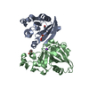
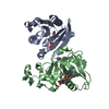
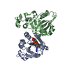
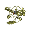
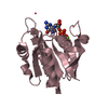
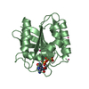
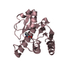
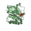
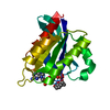
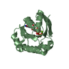
- PDB-2j5x: STRUCTURE OF THE SMALL G PROTEIN ARF6 IN COMPLEX WITH GTPGAMMAS -
+
Open data
ID or keywords:
Loading...
-
Basic information
Entry
Database: PDB / ID: 2j5x
Title
STRUCTURE OF THE SMALL G PROTEIN ARF6 IN COMPLEX WITH GTPGAMMAS
Components
ADP-RIBOSYLATION FACTOR 6
Keywords
PROTEIN TRANSPORT / MEMBRANE TRAFFIC / RAS / ARF / ARF6 / G PROTEIN / MYRISTATE / TRANSPORT / ER-GOLGI TRANSPORT / NUCLEOTIDE-BINDING / LIPOPROTEIN / GTP-BINDING / GOLGI APPARATUS
Function / homology
Function and homology information
erythrocyte apoptotic process / maintenance of postsynaptic density structure / protein localization to cleavage furrow / positive regulation of mitotic cytokinetic process / establishment of epithelial cell polarity / regulation of dendritic spine development / negative regulation of protein localization to cell surface / protein localization to endosome / regulation of Rac protein signal transduction / negative regulation of dendrite development ...erythrocyte apoptotic process / maintenance of postsynaptic density structure / protein localization to cleavage furrow / positive regulation of mitotic cytokinetic process / establishment of epithelial cell polarity / regulation of dendritic spine development / negative regulation of protein localization to cell surface / protein localization to endosome / regulation of Rac protein signal transduction / negative regulation of dendrite development / ruffle assembly / positive regulation of keratinocyte migration / regulation of filopodium assembly / positive regulation of focal adhesion disassembly / endocytic recycling / MET receptor recycling / thioesterase binding / Flemming body / filopodium membrane / TBC/RABGAPs / protein localization to cell surface / cortical actin cytoskeleton organization / hepatocyte apoptotic process / positive regulation of actin filament polymerization / cleavage furrow / endocytic vesicle / regulation of presynapse assembly / synaptic vesicle endocytosis / ruffle / vesicle-mediated transport / signaling adaptor activity / small monomeric GTPase / protein localization to plasma membrane / positive regulation of protein localization to plasma membrane / positive regulation of protein secretion / liver development / negative regulation of receptor-mediated endocytosis / cellular response to nerve growth factor stimulus / positive regulation of neuron projection development / intracellular protein transport / recycling endosome membrane / GDP binding / nervous system development / Clathrin-mediated endocytosis / presynapse / G protein activity / early endosome membrane / midbody / cell cortex / cell differentiation / cell adhesion / endosome / postsynapse / cell division / focal adhesion / GTPase activity / GTP binding / glutamatergic synapse / Golgi apparatus / extracellular exosome / membrane / plasma membrane / cytoplasm / cytosol Similarity search - Function
ADP-ribosylation factor 6 / Small GTPase superfamily, ARF type / Small GTPase Arf domain profile. / Sar1p-like members of the Ras-family of small GTPases / Small GTPase superfamily, ARF/SAR type / ADP-ribosylation factor family / ARF-like small GTPases; ARF, ADP-ribosylation factor / Rab subfamily of small GTPases / Small GTP-binding protein domain / P-loop containing nucleotide triphosphate hydrolases ...ADP-ribosylation factor 6 / Small GTPase superfamily, ARF type / Small GTPase Arf domain profile. / Sar1p-like members of the Ras-family of small GTPases / Small GTPase superfamily, ARF/SAR type / ADP-ribosylation factor family / ARF-like small GTPases; ARF, ADP-ribosylation factor / Rab subfamily of small GTPases / Small GTP-binding protein domain / P-loop containing nucleotide triphosphate hydrolases / Rossmann fold / P-loop containing nucleoside triphosphate hydrolase / 3-Layer(aba) Sandwich / Alpha Beta Similarity search - Domain/homology
Mass: 20110.166 Da / Num. of mol.: 2 Source method: isolated from a genetically manipulated source Source: (gene. exp.) HOMO SAPIENS (human) / Production host: ESCHERICHIA COLI (E. coli) / References: UniProt: P62330
Protocol: SINGLE WAVELENGTH / Monochromatic (M) / Laue (L): M / Scattering type: x-ray
Radiation wavelength
Wavelength: 0.934 Å / Relative weight: 1
Reflection
Resolution: 2.8→30 Å / Num. obs: 8616 / % possible obs: 93.1 % / Observed criterion σ(I): 2 / Redundancy: 6 % / Biso Wilson estimate: 94.5 Å2 / Rmerge(I) obs: 0.06 / Net I/σ(I): 25.2
Reflection shell
Resolution: 2.8→2.98 Å / % possible all: 83.6
-
Processing
Software
Name
Version
Classification
CNS
1
refinement
DENZO
datareduction
SCALEPACK
datascaling
AMoRE
phasing
Refinement
Method to determine structure: MOLECULAR REPLACEMENT Starting model: ARF1DELTA17-GDPNHP, FROM GOLDBERG J. Resolution: 2.8→29.97 Å / Rfactor Rfree error: 0.009 / Isotropic thermal model: RESTRAINED / Cross valid method: THROUGHOUT / σ(F): 0.5 Details: THE FIRST TEN RESIDUES IN CHAIN A AND B WERE NOT VISIBLE IN THE DENSITY MAPS.
In the structure databanks used in Yorodumi, some data are registered as the other names, "COVID-19 virus" and "2019-nCoV". Here are the details of the virus and the list of structure data.
Jan 31, 2019. EMDB accession codes are about to change! (news from PDBe EMDB page)
EMDB accession codes are about to change! (news from PDBe EMDB page)
The allocation of 4 digits for EMDB accession codes will soon come to an end. Whilst these codes will remain in use, new EMDB accession codes will include an additional digit and will expand incrementally as the available range of codes is exhausted. The current 4-digit format prefixed with “EMD-” (i.e. EMD-XXXX) will advance to a 5-digit format (i.e. EMD-XXXXX), and so on. It is currently estimated that the 4-digit codes will be depleted around Spring 2019, at which point the 5-digit format will come into force.
The EM Navigator/Yorodumi systems omit the EMD- prefix.
Related info.:Q: What is EMD? / ID/Accession-code notation in Yorodumi/EM Navigator
Yorodumi is a browser for structure data from EMDB, PDB, SASBDB, etc.
This page is also the successor to EM Navigator detail page, and also detail information page/front-end page for Omokage search.
The word "yorodu" (or yorozu) is an old Japanese word meaning "ten thousand". "mi" (miru) is to see.
Related info.:EMDB / PDB / SASBDB / Comparison of 3 databanks / Yorodumi Search / Aug 31, 2016. New EM Navigator & Yorodumi / Yorodumi Papers / Jmol/JSmol / Function and homology information / Changes in new EM Navigator and Yorodumi
 Movie
Movie Controller


 Open data
 Basic information
 Components
 Keywords
 Function and homology information
 HOMO SAPIENS (human)
 X-RAY DIFFRACTION /
  Authors
 Citation
 Structure visualization
 Downloads & links
 Other downloads
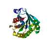


 PDBj
PDBj


 Assembly


 Mass: 539.246 Da / Num. of mol.: 2 / Source method: obtained synthetically / Formula: C10H16N5O13P3S
 Mass: 24.305 Da / Num. of mol.: 2 / Source method: obtained synthetically / Formula: Mg
 Mass: 18.015 Da / Num. of mol.: 32 / Source method: isolated from a natural source / Formula: H2O
 Sample preparation
 / Beamline: ID14-4 / Wavelength: 0.934
  Processing