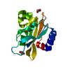
Entry Database : PDB / ID : 2j3pTitle crystal structure of rat FGF1 at 1.4 A HEPARIN-BINDING GROWTH FACTOR 1 Keywords / / / / / / / / Function / homology Function Domain/homology Component
/ / / / / / / / / / / / / / / / / / / / / / / / / / / / / / / / / / / / / / / / / / / / / / / / / / / / / / / / / / / / / / / / / / / / / / / / / / / / / / / / / / / / / / / / / / Biological species RATTUS NORVEGICUS (Norway rat)Method / / / Resolution : 1.4 Å Authors Kulahin, N. / Kristensen, O. / Berezin, V. / Gajhede, M. / Bock, E. Journal : Acta Crystallogr.,Sect.F / Year : 2007Title : Structure of Rat Acidic Fibroblast Growth Factor at 1.4 A Resolution.Authors : Kulahin, N. / Kiselyov, V. / Kochoyan, A. / Kristensen, O. / Kastrup, J.S. / Berezin, V. / Bock, E. / Gajhede, M. History Deposition Aug 22, 2006 Deposition site / Processing site Revision 1.0 Feb 13, 2007 Provider / Type Revision 1.1 May 8, 2011 Group Revision 1.2 Jul 13, 2011 Group Revision 1.3 Dec 13, 2023 Group Data collection / Database references ... Data collection / Database references / Other / Refinement description Category chem_comp_atom / chem_comp_bond ... chem_comp_atom / chem_comp_bond / database_2 / pdbx_database_status / pdbx_initial_refinement_model Item / _database_2.pdbx_database_accession / _pdbx_database_status.status_code_sf
Show all Show less
 Movie
Movie Controller


 Open data
 Basic information
 Components
 Keywords
 Function and homology information
 X-RAY DIFFRACTION /
  Authors
 Citation
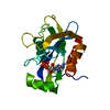
 Structure visualization
 Downloads & links
 Other downloads
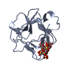


 PDBj
PDBj


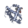
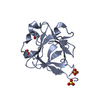
 Assembly
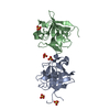


 Mass: 96.063 Da / Num. of mol.: 5 / Source method: obtained synthetically / Formula: SO4
 Mass: 18.015 Da / Num. of mol.: 255 / Source method: isolated from a natural source / Formula: H2O
 Sample preparation
 / Beamline: ID23-1 / Wavelength: 0.98
  Processing