 Movie
Movie Controller

[English] 日本語
 Yorodumi
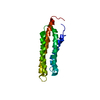
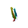
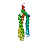
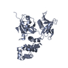
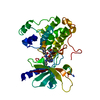
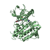
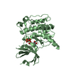
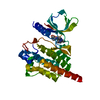
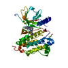
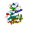
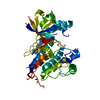
- PDB-2j0l: Crystal structure of a the active conformation of the kinase doma... -
+ Open data

- Basic information
| Entry | Database: PDB / ID: 2j0l | ||||||
|---|---|---|---|---|---|---|---|
| Title | Crystal structure of a the active conformation of the kinase domain of focal adhesion kinase with a phosphorylated activation loop. | ||||||
 Components Components | FOCAL ADHESION KINASE 1 | ||||||
 Keywords Keywords | TRANSFERASE / FOCAL ADHESION / CELL MIGRATION / PHOSPHORYLATION / FERM / KINASE / ATP-BINDING / INTEGRIN SIGNALING / NUCLEOTIDE-BINDING / TYROSINE-PROTEIN KINASE | ||||||
| Function / homology |  Function and homology information Function and homology informationradial glia-guided pyramidal neuron migration / negative regulation of protein autophosphorylation / positive regulation of protein tyrosine kinase activity / calcium-dependent cysteine-type endopeptidase activity / positive regulation of substrate-dependent cell migration, cell attachment to substrate / angiogenesis involved in wound healing / signal complex assembly / response to pH / wound healing, spreading of cells / positive regulation of protein binding ...radial glia-guided pyramidal neuron migration / negative regulation of protein autophosphorylation / positive regulation of protein tyrosine kinase activity / calcium-dependent cysteine-type endopeptidase activity / positive regulation of substrate-dependent cell migration, cell attachment to substrate / angiogenesis involved in wound healing / signal complex assembly / response to pH / wound healing, spreading of cells / positive regulation of protein binding / negative regulation of anoikis / negative regulation of cell-substrate adhesion / positive regulation of focal adhesion assembly / regulation of cell adhesion / response to muscle stretch / molecular function activator activity / actin filament organization / non-specific protein-tyrosine kinase / non-membrane spanning protein tyrosine kinase activity / sarcolemma / integrin binding / epidermal growth factor receptor signaling pathway / protein autophosphorylation / protease binding / protein tyrosine kinase activity / cell cortex / ciliary basal body / positive regulation of cell migration / focal adhesion / centrosome / positive regulation of cell population proliferation / perinuclear region of cytoplasm / ATP binding / identical protein binding / nucleus / plasma membrane / cytoplasm Similarity search - Function | ||||||
| Biological species |  | ||||||
| Method |  X-RAY DIFFRACTION / SYNCHROTRON / MOLECULAR REPLACEMENT / Resolution: 2.3 Å X-RAY DIFFRACTION / SYNCHROTRON / MOLECULAR REPLACEMENT / Resolution: 2.3 Å | ||||||
 Authors Authors | Lietha, D. / Cai, X. / Li, Y. / Schaller, M.D. / Eck, M.J. | ||||||
 Citation Citation | Journal: Cell(Cambridge,Mass.) / Year: 2007 Title: Structural Basis for the Autoinhibition of Focal Adhesion Kinase Authors: Lietha, D. / Cai, X. / Ceccarelli, D.F.J. / Li, Y. / Schaller, M.D. / Eck, M.J. | ||||||
| History |
|
- Structure visualization
| Structure viewer | Molecule: MolmilJmol/JSmol |
|---|
- Downloads & links
-Download
| PDBx/mmCIF format | 2j0l.cif.gz | 76.1 KB | Display | PDBx/mmCIF format |
|---|---|---|---|---|
| PDB format | pdb2j0l.ent.gz | 54.9 KB | Display | PDB format |
| PDBx/mmJSON format | 2j0l.json.gz | Tree view | PDBx/mmJSON format | |
| Others |  Other downloads Other downloads |
-Validation report
| Arichive directory | https://data.pdbj.org/pub/pdb/validation_reports/j0/2j0lftp://data.pdbj.org/pub/pdb/validation_reports/j0/2j0l | HTTPS FTP |
|---|
-Related structure data
| Related structure data | 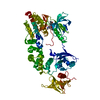 2j0jC  2j0kC  2j0mC 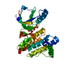 1mp8S C: citing same article ( S: Starting model for refinement |
|---|---|
| Similar structure data |


-Links
 PDBj
PDBj


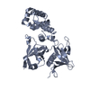
- Assembly
| Deposited unit | 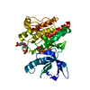
| ||||||||
|---|---|---|---|---|---|---|---|---|---|
| 1 |
| ||||||||
| Unit cell |
|
-Components
| #1: Protein | Mass: 31920.758 Da / Num. of mol.: 1 / Fragment: KINASE DOMAIN, RESIDUES 411-686 Source method: isolated from a genetically manipulated source Details: PHOSPHORYLATED ACTIVATION LOOP (Y576, Y577) / Source: (gene. exp.)  TRICHOPLUSIA NI (cabbage looper) TRICHOPLUSIA NI (cabbage looper)References: UniProt: Q00944, non-specific protein-tyrosine kinase |
|---|---|
| #2: Chemical | ChemComp-SO4 /   Mass: 96.063 Da / Num. of mol.: 1 / Source method: obtained synthetically / Formula: SO4 Mass: 96.063 Da / Num. of mol.: 1 / Source method: obtained synthetically / Formula: SO4 |
| #3: Chemical | ChemComp-MG /   Mass: 24.305 Da / Num. of mol.: 1 / Source method: obtained synthetically / Formula: Mg Mass: 24.305 Da / Num. of mol.: 1 / Source method: obtained synthetically / Formula: Mg |
| #4: Chemical | ChemComp-ANP /   Mass: 506.196 Da / Num. of mol.: 1 / Source method: obtained synthetically / Formula: C10H17N6O12P3 / Comment: AMP-PNP, energy-carrying molecule analogue*YM Mass: 506.196 Da / Num. of mol.: 1 / Source method: obtained synthetically / Formula: C10H17N6O12P3 / Comment: AMP-PNP, energy-carrying molecule analogue*YM |
| #5: Water | ChemComp-HOH /  Mass: 18.015 Da / Num. of mol.: 97 / Source method: isolated from a natural source / Formula: H2O Mass: 18.015 Da / Num. of mol.: 97 / Source method: isolated from a natural source / Formula: H2O |
| Has protein modification | Y |
| Sequence details | RESIDUES 411-686 |
-Experimental details
-Experiment
| Experiment | Method: X-RAY DIFFRACTION / Number of used crystals: 1 |
|---|
- Sample preparation
| Crystal | Density Matthews: 2.23 Å3/Da / Density % sol: 44.9 % |
|---|---|
| Crystal grow | pH: 8.5 Details: 26% PEG4K, 0.2M LISO4, 0.1M TRIS PH8.5, 10MM TCEP, pH 8.50 |
-Data collection
| Diffraction | Mean temperature: 100 K |
|---|---|
| Diffraction source | Source: SYNCHROTRON / Site: NSLS  / Beamline: X29A / Wavelength: 1.1 / Beamline: X29A / Wavelength: 1.1 |
| Detector | Type: ADSC CCD / Detector: CCD / Date: Sep 17, 2005 |
| Radiation | Protocol: SINGLE WAVELENGTH / Monochromatic (M) / Laue (L): M / Scattering type: x-ray |
| Radiation wavelength | Wavelength: 1.1 Å / Relative weight: 1 |
| Reflection | Resolution: 2.3→50 Å / Num. obs: 11899 / % possible obs: 92.3 % / Observed criterion σ(I): 0 / Redundancy: 4.1 % / Rmerge(I) obs: 0.1 / Net I/σ(I): 12.1 |
| Reflection shell | Resolution: 2.3→2.38 Å / Redundancy: 2.3 % / Rmerge(I) obs: 0.32 / Mean I/σ(I) obs: 2.51 / % possible all: 56.1 |
- Processing
| Software |
| ||||||||||||||||||||||||||||||||||||||||||||||||||||||||||||||||||||||||||||||||||||||||||||||||||||||||||||||||||||||||||||||||||||||||||||||||||||||||||||||||||||||||||||||||||||||
|---|---|---|---|---|---|---|---|---|---|---|---|---|---|---|---|---|---|---|---|---|---|---|---|---|---|---|---|---|---|---|---|---|---|---|---|---|---|---|---|---|---|---|---|---|---|---|---|---|---|---|---|---|---|---|---|---|---|---|---|---|---|---|---|---|---|---|---|---|---|---|---|---|---|---|---|---|---|---|---|---|---|---|---|---|---|---|---|---|---|---|---|---|---|---|---|---|---|---|---|---|---|---|---|---|---|---|---|---|---|---|---|---|---|---|---|---|---|---|---|---|---|---|---|---|---|---|---|---|---|---|---|---|---|---|---|---|---|---|---|---|---|---|---|---|---|---|---|---|---|---|---|---|---|---|---|---|---|---|---|---|---|---|---|---|---|---|---|---|---|---|---|---|---|---|---|---|---|---|---|---|---|---|---|
| Refinement | Method to determine structure: MOLECULAR REPLACEMENT Starting model: PDB ENTRY 1MP8 Resolution: 2.3→43.48 Å / Cor.coef. Fo:Fc: 0.937 / Cor.coef. Fo:Fc free: 0.912 / SU B: 15.316 / SU ML: 0.216 / TLS residual ADP flag: LIKELY RESIDUAL / Cross valid method: THROUGHOUT / ESU R: 0.543 / ESU R Free: 0.285 / Stereochemistry target values: MAXIMUM LIKELIHOOD / Details: HYDROGENS HAVE BEEN ADDED IN THE RIDING POSITIONS
| ||||||||||||||||||||||||||||||||||||||||||||||||||||||||||||||||||||||||||||||||||||||||||||||||||||||||||||||||||||||||||||||||||||||||||||||||||||||||||||||||||||||||||||||||||||||
| Solvent computation | Ion probe radii: 0.8 Å / Shrinkage radii: 0.8 Å / VDW probe radii: 1.4 Å / Solvent model: MASK | ||||||||||||||||||||||||||||||||||||||||||||||||||||||||||||||||||||||||||||||||||||||||||||||||||||||||||||||||||||||||||||||||||||||||||||||||||||||||||||||||||||||||||||||||||||||
| Displacement parameters | Biso mean: 39.27 Å2
| ||||||||||||||||||||||||||||||||||||||||||||||||||||||||||||||||||||||||||||||||||||||||||||||||||||||||||||||||||||||||||||||||||||||||||||||||||||||||||||||||||||||||||||||||||||||
| Refinement step | Cycle: LAST / Resolution: 2.3→43.48 Å
| ||||||||||||||||||||||||||||||||||||||||||||||||||||||||||||||||||||||||||||||||||||||||||||||||||||||||||||||||||||||||||||||||||||||||||||||||||||||||||||||||||||||||||||||||||||||
| Refine LS restraints |
|