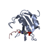
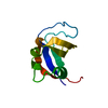
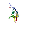
SHEET THE SHEET STRUCTURE OF THIS MOLECULE IS BIFURCATED. IN ORDER TO REPRESENT THIS FEATURE IN ... SHEET THE SHEET STRUCTURE OF THIS MOLECULE IS BIFURCATED. IN ORDER TO REPRESENT THIS FEATURE IN THE SHEET RECORDS BELOW, TWO SHEETS ARE DEFINED.
Mass: 18.015 Da / Num. of mol.: 101 / Source method: isolated from a natural source / Formula: H2O
Compound details
BINDS TO ACTIVATED (PHOSPHORYLATED) PROTEIN-TYR KINASES, THROUGH ITS SH2 DOMAIN, AND ACTS AS AN ...BINDS TO ACTIVATED (PHOSPHORYLATED) PROTEIN-TYR KINASES, THROUGH ITS SH2 DOMAIN, AND ACTS AS AN ADAPTER, MEDIATING THE ASSOCIATION OF THE P110 CATALYTIC UNIT TO THE PLASMA MEMBRANE. NECESSARY FOR THE INSULIN-STIMULATED INCREASE IN GLUCOSE UPTAKE AND GLYCOGEN SYNTHESIS IN INSULIN-SENSITIVE TISSUES.
Protocol: SINGLE WAVELENGTH / Monochromatic (M) / Laue (L): M / Scattering type: x-ray
Radiation wavelength
Wavelength: 1.5418 Å / Relative weight: 1
Reflection
Resolution: 2→20 Å / Num. obs: 8651 / % possible obs: 96.5 % / Rmerge(I) obs: 0.05 / Net I/σ(I): 30.87
Reflection shell
Resolution: 2→2.1 Å / Rmerge(I) obs: 0.14 / Mean I/σ(I) obs: 10.84 / % possible all: 92.7
-
Processing
Software
Name: X-PLOR / Classification: refinement
Refinement
Method to determine structure: OTHER / Resolution: 2→15 Å / Cross valid method: THROUGHOUT / σ(F): 0 Details: THIS COODINATES FILE WAS DEPOSITED 10 YEARS AFTER THE PUBLICATION OF THE WORK. SOME OF THE DATA ARE MISSING AND ARE SHOWN AS '00000'
Rfactor
% reflection
Rfree
0.245
-
Rwork
0.1539
-
obs
0.1539
0 %
Displacement parameters
Biso mean: 17.65 Å2
Refinement step
Cycle: LAST / Resolution: 2→15 Å
Protein
Nucleic acid
Ligand
Solvent
Total
Num. atoms
1018
0
0
101
1119
Refine LS restraints
Refine-ID
Type
Dev ideal
X-RAY DIFFRACTION
x_bond_d
0.01
X-RAY DIFFRACTION
x_bond_d_na
X-RAY DIFFRACTION
x_bond_d_prot
X-RAY DIFFRACTION
x_angle_d
X-RAY DIFFRACTION
x_angle_d_na
X-RAY DIFFRACTION
x_angle_d_prot
X-RAY DIFFRACTION
x_angle_deg
1.728
X-RAY DIFFRACTION
x_angle_deg_na
X-RAY DIFFRACTION
x_angle_deg_prot
X-RAY DIFFRACTION
x_dihedral_angle_d
X-RAY DIFFRACTION
x_dihedral_angle_d_na
X-RAY DIFFRACTION
x_dihedral_angle_d_prot
X-RAY DIFFRACTION
x_improper_angle_d
X-RAY DIFFRACTION
x_improper_angle_d_na
X-RAY DIFFRACTION
x_improper_angle_d_prot
X-RAY DIFFRACTION
x_mcbond_it
X-RAY DIFFRACTION
x_mcangle_it
X-RAY DIFFRACTION
x_scbond_it
X-RAY DIFFRACTION
x_scangle_it
+
About Yorodumi
-
News
-
Feb 9, 2022. New format data for meta-information of EMDB entries
New format data for meta-information of EMDB entries
Version 3 of the EMDB header file is now the official format.
The previous official version 1.9 will be removed from the archive.
In the structure databanks used in Yorodumi, some data are registered as the other names, "COVID-19 virus" and "2019-nCoV". Here are the details of the virus and the list of structure data.
Jan 31, 2019. EMDB accession codes are about to change! (news from PDBe EMDB page)
EMDB accession codes are about to change! (news from PDBe EMDB page)
The allocation of 4 digits for EMDB accession codes will soon come to an end. Whilst these codes will remain in use, new EMDB accession codes will include an additional digit and will expand incrementally as the available range of codes is exhausted. The current 4-digit format prefixed with “EMD-” (i.e. EMD-XXXX) will advance to a 5-digit format (i.e. EMD-XXXXX), and so on. It is currently estimated that the 4-digit codes will be depleted around Spring 2019, at which point the 5-digit format will come into force.
The EM Navigator/Yorodumi systems omit the EMD- prefix.
Related info.:Q: What is EMD? / ID/Accession-code notation in Yorodumi/EM Navigator
Yorodumi is a browser for structure data from EMDB, PDB, SASBDB, etc.
This page is also the successor to EM Navigator detail page, and also detail information page/front-end page for Omokage search.
The word "yorodu" (or yorozu) is an old Japanese word meaning "ten thousand". "mi" (miru) is to see.
Related info.:EMDB / PDB / SASBDB / Comparison of 3 databanks / Yorodumi Search / Aug 31, 2016. New EM Navigator & Yorodumi / Yorodumi Papers / Jmol/JSmol / Function and homology information / Changes in new EM Navigator and Yorodumi
 Movie
Movie Controller

 Yorodumi
 Open data
 Basic information
 Components
 Keywords
 Function and homology information
 HOMO SAPIENS (human)
 X-RAY DIFFRACTION / OTHER / Resolution: 2 Å
  Authors
 Citation
 Structure visualization
 Downloads & links
 Other downloads
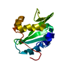


 PDBj
PDBj


 Assembly

 Mass: 18.015 Da / Num. of mol.: 101 / Source method: isolated from a natural source / Formula: H2O
 Sample preparation
 Processing