 Movie
Movie Controller

[English] 日本語
 Yorodumi
- PDB-2itm: Crystal structure of the E. coli xylulose kinase complexed with x... -
+ Open data

- Basic information
| Entry | Database: PDB / ID: 2itm | ||||||
|---|---|---|---|---|---|---|---|
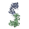
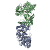
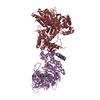
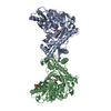
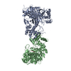
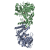
| Title | Crystal structure of the E. coli xylulose kinase complexed with xylulose | ||||||
 Components Components | Xylulose kinase | ||||||
 Keywords Keywords | TRANSFERASE / xylulokinase / xylulose / kinase / ATPase / FGGY kinase | ||||||
| Function / homology |  Function and homology information Function and homology information1-deoxy-D-xylulose kinase activity / xylulokinase / xylulose catabolic process / D-xylulokinase activity / D-xylose catabolic process / Transferases; Transferring phosphorus-containing groups; Phosphotransferases with an alcohol group as acceptor / kinase activity / protein homodimerization activity / ATP binding Similarity search - Function | ||||||
| Biological species |  | ||||||
| Method |  X-RAY DIFFRACTION / SYNCHROTRON / MIR / Resolution: 2.1 Å X-RAY DIFFRACTION / SYNCHROTRON / MIR / Resolution: 2.1 Å | ||||||
 Authors Authors | di Luccio, E. / Voegtli, J. / Wilson, D.K. | ||||||
 Citation Citation | Journal: J.Mol.Biol. / Year: 2007 Title: Structural and kinetic studies of induced fit in xylulose kinase from Escherichia coli. Authors: Di Luccio, E. / Petschacher, B. / Voegtli, J. / Chou, H.T. / Stahlberg, H. / Nidetzky, B. / Wilson, D.K. | ||||||
| History |
|
- Structure visualization
| Structure viewer | Molecule: MolmilJmol/JSmol |
|---|
- Downloads & links
-Download
| PDBx/mmCIF format | 2itm.cif.gz | 202.3 KB | Display | PDBx/mmCIF format |
|---|---|---|---|---|
| PDB format | pdb2itm.ent.gz | 161.4 KB | Display | PDB format |
| PDBx/mmJSON format | 2itm.json.gz | Tree view | PDBx/mmJSON format | |
| Others |  Other downloads Other downloads |
-Validation report
| Arichive directory | https://data.pdbj.org/pub/pdb/validation_reports/it/2itmftp://data.pdbj.org/pub/pdb/validation_reports/it/2itm | HTTPS FTP |
|---|
-Related structure data

-Links
 PDBj
PDBj
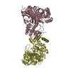
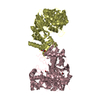
- Assembly
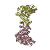
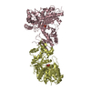
| Deposited unit | 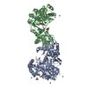
| |||||||||||||||||||||||||||||||||||||||||||||||||||||||||||||||||||||||||||||||||||||||||||||||||||||||||||||||||||||||||||||||||||||||||||||||||||||||||||||||||||||||||||||||||||||||||||||||||||
|---|---|---|---|---|---|---|---|---|---|---|---|---|---|---|---|---|---|---|---|---|---|---|---|---|---|---|---|---|---|---|---|---|---|---|---|---|---|---|---|---|---|---|---|---|---|---|---|---|---|---|---|---|---|---|---|---|---|---|---|---|---|---|---|---|---|---|---|---|---|---|---|---|---|---|---|---|---|---|---|---|---|---|---|---|---|---|---|---|---|---|---|---|---|---|---|---|---|---|---|---|---|---|---|---|---|---|---|---|---|---|---|---|---|---|---|---|---|---|---|---|---|---|---|---|---|---|---|---|---|---|---|---|---|---|---|---|---|---|---|---|---|---|---|---|---|---|---|---|---|---|---|---|---|---|---|---|---|---|---|---|---|---|---|---|---|---|---|---|---|---|---|---|---|---|---|---|---|---|---|---|---|---|---|---|---|---|---|---|---|---|---|---|---|---|---|---|
| 1 |
| |||||||||||||||||||||||||||||||||||||||||||||||||||||||||||||||||||||||||||||||||||||||||||||||||||||||||||||||||||||||||||||||||||||||||||||||||||||||||||||||||||||||||||||||||||||||||||||||||||
| Unit cell |
| |||||||||||||||||||||||||||||||||||||||||||||||||||||||||||||||||||||||||||||||||||||||||||||||||||||||||||||||||||||||||||||||||||||||||||||||||||||||||||||||||||||||||||||||||||||||||||||||||||
| Noncrystallographic symmetry (NCS) | NCS domain:
NCS domain segments: Ens-ID: 1 / Refine code: 3
|