 Movie
Movie Controller


+ Open data

- Basic information
| Entry | Database: PDB / ID: 2ill | ||||||
|---|---|---|---|---|---|---|---|
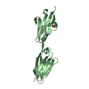
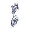
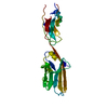
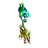
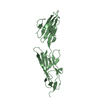
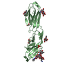
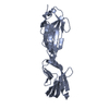
| Title | Anomalous substructure of Titin-A168169 | ||||||
 Components Components | Titin | ||||||
 Keywords Keywords | TRANSFERASE / long wavelength | ||||||
| Function / homology |  Function and homology information Function and homology informationsarcomerogenesis / titin-telethonin complex / structural molecule activity conferring elasticity / skeletal muscle myosin thick filament assembly / telethonin binding / detection of muscle stretch / : / muscle alpha-actinin binding / cardiac myofibril assembly / cardiac muscle hypertrophy ...sarcomerogenesis / titin-telethonin complex / structural molecule activity conferring elasticity / skeletal muscle myosin thick filament assembly / telethonin binding / detection of muscle stretch / : / muscle alpha-actinin binding / cardiac myofibril assembly / cardiac muscle hypertrophy / cardiac muscle tissue morphogenesis / protein kinase regulator activity / mitotic chromosome condensation / Striated Muscle Contraction / muscle filament sliding / actinin binding / M band / cardiac muscle cell development / I band / sarcomere organization / structural constituent of muscle / striated muscle thin filament / skeletal muscle thin filament assembly / striated muscle contraction / skeletal muscle contraction / cardiac muscle contraction / muscle contraction / condensed nuclear chromosome / positive regulation of protein secretion / response to calcium ion / Z disc / actin filament binding / Platelet degranulation / actin cytoskeleton / protein tyrosine kinase activity / protease binding / calmodulin binding / non-specific serine/threonine protein kinase / protein serine kinase activity / protein serine/threonine kinase activity / calcium ion binding / positive regulation of gene expression / protein kinase binding / enzyme binding / protein homodimerization activity / extracellular exosome / extracellular region / ATP binding / identical protein binding / cytosol Similarity search - Function | ||||||
| Biological species |  Homo sapiens (human) Homo sapiens (human) | ||||||
| Method |  X-RAY DIFFRACTION / SYNCHROTRON / FOURIER SYNTHESIS / Resolution: 2.2 Å X-RAY DIFFRACTION / SYNCHROTRON / FOURIER SYNTHESIS / Resolution: 2.2 Å | ||||||
 Authors Authors | Mueller-Dieckmann, C. / Weiss, M.S. | ||||||
 Citation Citation | Journal: ACTA CRYSTALLOGR.,SECT.D / Year: 2007 Title: On the routine use of soft X-rays in macromolecular crystallography. Part IV. Efficient determination of anomalous substructures in biomacromolecules using longer X-ray wavelengths Authors: Mueller-Dieckmann, C. / Panjikar, S. / Schmidt, A. / Mueller, S. / Kuper, J. / Geerlof, A. / Wilmanns, M. / Singh, R.K. / Tucker, P.A. / Weiss, M.S. | ||||||
| History |
|
- Structure visualization
| Structure viewer | Molecule: MolmilJmol/JSmol |
|---|
- Downloads & links
-Download
| PDBx/mmCIF format | 2ill.cif.gz | 53.5 KB | Display | PDBx/mmCIF format |
|---|---|---|---|---|
| PDB format | pdb2ill.ent.gz | 38.7 KB | Display | PDB format |
| PDBx/mmJSON format | 2ill.json.gz | Tree view | PDBx/mmJSON format | |
| Others |  Other downloads Other downloads |
-Validation report
| Arichive directory | https://data.pdbj.org/pub/pdb/validation_reports/il/2illftp://data.pdbj.org/pub/pdb/validation_reports/il/2ill | HTTPS FTP |
|---|
-Related structure data
| Related structure data |  2g4hC 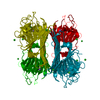 2g4iC 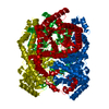 2g4jC 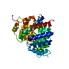 2g4kC 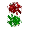 2g4lC  2g4mC  2g4nC  2g4oC 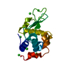 2g4pC 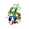 2g4qC 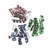 2g4rC  2g4sC 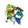 2g4tC  2g4uC 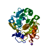 2g4vC 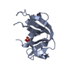 2g4wC 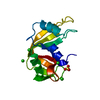 2g4xC 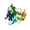 2g4yC 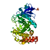 2g4zC 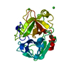 2g51C 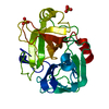 2g52C 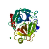 2g55C C: citing same article ( |
|---|---|
| Similar structure data |


-Links
 PDBj
PDBj


- Assembly
| Deposited unit | 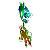
| ||||||||
|---|---|---|---|---|---|---|---|---|---|
| 1 |
| ||||||||
| Unit cell |
|
-Components
| #1: Protein | Mass: 21637.691 Da / Num. of mol.: 1 / Fragment: Titin-A168169 Source method: isolated from a genetically manipulated source Source: (gene. exp.) Homo sapiens (human) / Production host:  References: UniProt: Q8WZ42, non-specific serine/threonine protein kinase | ||
|---|---|---|---|
| #2: Chemical |   Mass: 35.453 Da / Num. of mol.: 2 / Source method: obtained synthetically / Formula: Cl Mass: 35.453 Da / Num. of mol.: 2 / Source method: obtained synthetically / Formula: Cl#3: Water | ChemComp-HOH / |  Mass: 18.015 Da / Num. of mol.: 94 / Source method: isolated from a natural source / Formula: H2O Mass: 18.015 Da / Num. of mol.: 94 / Source method: isolated from a natural source / Formula: H2O |
-Experimental details
-Experiment
| Experiment | Method: X-RAY DIFFRACTION / Number of used crystals: 1 |
|---|
- Sample preparation
| Crystal | Density Matthews: 3.69 Å3/Da / Density % sol: 66.67 % |
|---|
-Data collection
| Diffraction | Mean temperature: 100 K |
|---|---|
| Diffraction source | Source: SYNCHROTRON / Site: EMBL/DESY, HAMBURG  / Beamline: X12 / Wavelength: 2 Å / Beamline: X12 / Wavelength: 2 Å |
| Detector | Type: MAR CCD 165 mm / Detector: CCD |
| Radiation | Protocol: SINGLE WAVELENGTH / Monochromatic (M) / Laue (L): M / Scattering type: x-ray |
| Radiation wavelength | Wavelength: 2 Å / Relative weight: 1 |
| Reflection | Resolution: 2.2→99 Å / Num. all: 16570 / Num. obs: 16486 / % possible obs: 99 % / Redundancy: 11.8 % / Biso Wilson estimate: 47.5 Å2 / Rmerge(I) obs: 0.057 / Net I/σ(I): 34.1 |
| Reflection shell | Resolution: 2.2→2.26 Å / Redundancy: 10.3 % / Rmerge(I) obs: 0.45 / Mean I/σ(I) obs: 4.3 / % possible all: 92 |
- Processing
| Software |
| |||||||||||||||||||||||||||||||||||||||||||||||||||||||||||||||||||||||||||||||||||||||||||||||||||||||||||||||||||||||||||||
|---|---|---|---|---|---|---|---|---|---|---|---|---|---|---|---|---|---|---|---|---|---|---|---|---|---|---|---|---|---|---|---|---|---|---|---|---|---|---|---|---|---|---|---|---|---|---|---|---|---|---|---|---|---|---|---|---|---|---|---|---|---|---|---|---|---|---|---|---|---|---|---|---|---|---|---|---|---|---|---|---|---|---|---|---|---|---|---|---|---|---|---|---|---|---|---|---|---|---|---|---|---|---|---|---|---|---|---|---|---|---|---|---|---|---|---|---|---|---|---|---|---|---|---|---|---|---|
| Refinement | Method to determine structure: FOURIER SYNTHESIS / Resolution: 2.2→30 Å / Cor.coef. Fo:Fc: 0.944 / Cor.coef. Fo:Fc free: 0.92 / SU B: 8.818 / SU ML: 0.118 / TLS residual ADP flag: LIKELY RESIDUAL / Cross valid method: THROUGHOUT / ESU R: 0.193 / ESU R Free: 0.171 / Stereochemistry target values: MAXIMUM LIKELIHOOD / Details: HYDROGENS HAVE BEEN ADDED IN THE RIDING POSITIONS
| |||||||||||||||||||||||||||||||||||||||||||||||||||||||||||||||||||||||||||||||||||||||||||||||||||||||||||||||||||||||||||||
| Solvent computation | Ion probe radii: 0.8 Å / Shrinkage radii: 0.8 Å / VDW probe radii: 1.4 Å / Solvent model: BABINET MODEL WITH MASK | |||||||||||||||||||||||||||||||||||||||||||||||||||||||||||||||||||||||||||||||||||||||||||||||||||||||||||||||||||||||||||||
| Displacement parameters | Biso mean: 42.868 Å2
| |||||||||||||||||||||||||||||||||||||||||||||||||||||||||||||||||||||||||||||||||||||||||||||||||||||||||||||||||||||||||||||
| Refinement step | Cycle: LAST / Resolution: 2.2→30 Å
| |||||||||||||||||||||||||||||||||||||||||||||||||||||||||||||||||||||||||||||||||||||||||||||||||||||||||||||||||||||||||||||
| Refine LS restraints |
| |||||||||||||||||||||||||||||||||||||||||||||||||||||||||||||||||||||||||||||||||||||||||||||||||||||||||||||||||||||||||||||
| LS refinement shell | Resolution: 2.2→2.257 Å / Total num. of bins used: 20
| |||||||||||||||||||||||||||||||||||||||||||||||||||||||||||||||||||||||||||||||||||||||||||||||||||||||||||||||||||||||||||||
| Refinement TLS params. | Method: refined / Origin x: 11.0123 Å / Origin y: 56.0772 Å / Origin z: -6.4602 Å
|
