 Movie
Movie Controller

[English] 日本語
 Yorodumi
- PDB-2iif: single chain Integration Host Factor mutant protein (scIHF2-K45aE... -
+ Open data

- Basic information
| Entry | Database: PDB / ID: 2iif | ||||||
|---|---|---|---|---|---|---|---|
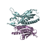
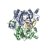
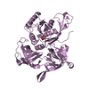
| Title | single chain Integration Host Factor mutant protein (scIHF2-K45aE) in complex with DNA | ||||||
 Components Components |
| ||||||
 Keywords Keywords | RECOMBINATION/DNA / DNA Kinking / bending / U-turn / intercalation / divalent / RECOMBINATION-DNA COMPLEX | ||||||
| Function / homology |  Function and homology information Function and homology informationIHF-DNA complex / DNA-binding transcription activator activity / protein-DNA complex / structural constituent of chromatin / regulation of translation / chromosome / DNA recombination / DNA replication / transcription cis-regulatory region binding / regulation of DNA-templated transcription ...IHF-DNA complex / DNA-binding transcription activator activity / protein-DNA complex / structural constituent of chromatin / regulation of translation / chromosome / DNA recombination / DNA replication / transcription cis-regulatory region binding / regulation of DNA-templated transcription / DNA-templated transcription / DNA binding / cytosol Similarity search - Function | ||||||
| Biological species |  synthetic construct (others) | ||||||
| Method |  X-RAY DIFFRACTION / FOURIER SYNTHESIS / Resolution: 2.72 Å X-RAY DIFFRACTION / FOURIER SYNTHESIS / Resolution: 2.72 Å | ||||||
 Authors Authors | Bao, Q. / Droege, P. / Davey, C.A. | ||||||
 Citation Citation | Journal: J.Mol.Biol. / Year: 2007 Title: A Divalent Metal-mediated Switch Controlling Protein-induced DNA Bending Authors: Bao, Q. / Chen, H. / Liu, Y. / Yan, J. / Droge, P. / Davey, C.A. #1: Journal: Nucleic Acids Res. / Year: 2003 Title: Activation of site-specific DNA integration in human cells by a single chain integration host factor Authors: Corona, T. / Bao, Q. / Christ, N. / Schwartz, T. / Li, J. / Droege, P. #2: Journal: Gene / Year: 2004 Title: Single-chain integration host factors as probes for high-precision nucleoprotein complex formation Authors: Bao, Q. / Christ, N. / Droege, P. | ||||||
| History |
|
- Structure visualization
| Structure viewer | Molecule: MolmilJmol/JSmol |
|---|
- Downloads & links
-Download
| PDBx/mmCIF format | 2iif.cif.gz | 98.3 KB | Display | PDBx/mmCIF format |
|---|---|---|---|---|
| PDB format | pdb2iif.ent.gz | 69.9 KB | Display | PDB format |
| PDBx/mmJSON format | 2iif.json.gz | Tree view | PDBx/mmJSON format | |
| Others |  Other downloads Other downloads |
-Validation report
| Arichive directory | https://data.pdbj.org/pub/pdb/validation_reports/ii/2iifftp://data.pdbj.org/pub/pdb/validation_reports/ii/2iif | HTTPS FTP |
|---|
-Related structure data


-Links
 PDBj
PDBj


- Assembly
| Deposited unit | 
| ||||||||
|---|---|---|---|---|---|---|---|---|---|
| 1 |
| ||||||||
| Unit cell |
|
-Components
-DNA chain , 3 types, 3 molecules CDE
| #1: DNA chain | Mass: 10801.969 Da / Num. of mol.: 1 / Source method: obtained synthetically / Details: chemically synthesized / Source: (synth.) synthetic construct (others) |
|---|---|
| #2: DNA chain | Mass: 4611.039 Da / Num. of mol.: 1 / Source method: obtained synthetically / Details: chemically synthesized / Source: (synth.) synthetic construct (others) |
| #3: DNA chain | Mass: 6074.938 Da / Num. of mol.: 1 / Source method: obtained synthetically / Details: chemically synthesized / Source: (synth.) synthetic construct (others) |
-Protein , 1 types, 1 molecules A
| #4: Protein | Mass: 22884.816 Da / Num. of mol.: 1 / Mutation: K89E Source method: isolated from a genetically manipulated source Source: (gene. exp.) |
|---|
-Non-polymers , 2 types, 171 molecules 

| #5: Chemical | ChemComp-MN /  Mass: 54.938 Da / Num. of mol.: 8 / Source method: obtained synthetically / Formula: Mn Mass: 54.938 Da / Num. of mol.: 8 / Source method: obtained synthetically / Formula: Mn#6: Water | ChemComp-HOH / | Mass: 18.015 Da / Num. of mol.: 163 / Source method: isolated from a natural source / Formula: H2O |
|---|
-Experimental details
-Experiment
| Experiment | Method: X-RAY DIFFRACTION / Number of used crystals: 1 |
|---|
- Sample preparation
| Crystal | Density Matthews: 2.58 Å3/Da / Density % sol: 52.28 % | ||||||||||||||||||||||||||||||||||||||||||||
|---|---|---|---|---|---|---|---|---|---|---|---|---|---|---|---|---|---|---|---|---|---|---|---|---|---|---|---|---|---|---|---|---|---|---|---|---|---|---|---|---|---|---|---|---|---|
| Crystal grow | Temperature: 291 K / Method: vapor diffusion, hanging drop / pH: 7.5 Details: 1:1 mix water/ well solution containing: 50mM Tris(pH 7.5), 25% PEG5000-MME, 20% glycerol, 50mM NaCl, 7.5mM MnCl2, VAPOR DIFFUSION, HANGING DROP, temperature 291K | ||||||||||||||||||||||||||||||||||||||||||||
| Components of the solutions |
|
-Data collection
| Diffraction | Mean temperature: 103 K |
|---|---|
| Diffraction source | Source: ROTATING ANODE / Type: RIGAKU / Wavelength: 1.542 Å |
| Detector | Type: RIGAKU RAXIS IV / Detector: IMAGE PLATE / Date: Oct 1, 2005 |
| Radiation | Monochromator: graphite / Protocol: SINGLE WAVELENGTH / Monochromatic (M) / Laue (L): M / Scattering type: x-ray |
| Radiation wavelength | Wavelength: 1.542 Å / Relative weight: 1 |
| Reflection | Resolution: 2.72→45 Å / Num. all: 12904 / Num. obs: 12530 / % possible obs: 97.1 % / Observed criterion σ(F): 0 / Observed criterion σ(I): 0 |
| Reflection shell | Resolution: 2.72→2.87 Å / % possible all: 95.9 |
- Processing
| Software |
| ||||||||||||||||||||||||||||
|---|---|---|---|---|---|---|---|---|---|---|---|---|---|---|---|---|---|---|---|---|---|---|---|---|---|---|---|---|---|
| Refinement | Method to determine structure: FOURIER SYNTHESIS / Resolution: 2.72→40 Å / Cross valid method: THROUGHOUT / σ(F): 0 / Stereochemistry target values: Engh & Huber
| ||||||||||||||||||||||||||||
| Solvent computation | Bsol: 29.174 Å2 | ||||||||||||||||||||||||||||
| Displacement parameters | Biso mean: 52.478 Å2 | ||||||||||||||||||||||||||||
| Refinement step | Cycle: LAST / Resolution: 2.72→40 Å
| ||||||||||||||||||||||||||||
| Refine LS restraints |
| ||||||||||||||||||||||||||||
| Xplor file |
|
